Figures & data
Figure 1 (A and B) Grey scale ultrasound pictures. (A) Longitudinal view showing a normal 4.5 mm thick Achilles tendon midportion. (B) Cross view showing a plantaris tendon (marked) localised close to the medial side of a normal Achilles tendon midportion.
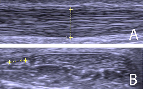
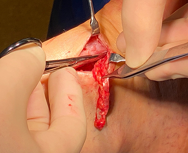
Figure 2 Picture from a patient with Plantaris tendinopathy alone, where 5–6 cm of a thickened plantaris tendon embedded in fat tissue is excised and removed from the medial side of a normal Achilles tendon midportion.