Figures & data
Figure 1 Electrocardiography showing bi-fascicular block.
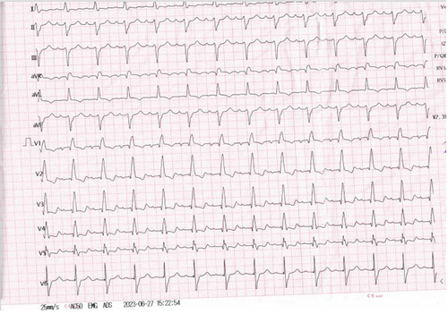
Figure 2 (A) Parasternal short axis view sinus Valsalva aneurysm dissecting in to interventricular septum (red arrow), (B) Apical 4 chamber view interventricular aneurysm (red arrow).
Abbreviations: LA, left atrium; LV, left ventricular; SVA, sinus Valsalva aneurysm; RV, right ventricular.
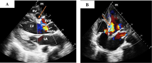
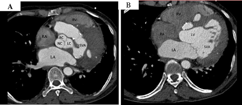
Figure 3 Cardiac CT in axial view (A and B) Showing left sinus of Valsalva aneurysm with suspicious communication in to interventricular septum.
Abbreviations: CT, computed tomography; RV, right ventricle; RA, right atrium; LA, left atrium; RC, right coronary sinus; LC, left coronary sinus; NC, non-coronary sinus; SVA, sinus valsalva aneurysm; LV, left ventricle.