
Figures & data
Figure 1 MR-HIFU treatment planning with superimposed MRI in three planes, such as coronal (A), sagittal (B), and transversal (C); pretreatment T2-weighted planning MR images show a uterine fibroid with low signal intensity. Red circles indicate the target volume for HIFU treatment as defined by the borders of the UF with a safety margin; green ellipsoids indicate the anticipated volumes of sonication.
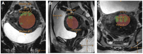
Figure 2 Sagittal T2-weighted MRI for planning purposes (A) shows a homogeneously hypointense UF in the anterior wall. Sagittal T1-weighted, fat-saturated and contrast-enhanced MRI immediately (B) as well as 1 month (C) after the MR-HIFU procedure with a satisfying NPV.
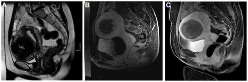
Table 1 Indications for MR-HIFU therapy of UFs
Table 2 Contraindications for MR-HIFU treatment of UFs
Table 3 Adverse effects and complications after MR-HIFU therapy of UFs