
Figures & data
Figure 1 A representative scheme of the lipids and EA comprising UDVs and the carried molecules.
Note: Copyright © 2013. Dove Medical Press. Adapted from Romero EL, Morilla MJ. Highly deformable and highly fluid vesicles as potential drug delivery systems: theoretical and practical considerations. Int J Nanomedicine. 2013;8:3171–3186.Citation10
Abbreviations: EA, edge activator; UDV, ultradeformable vesicle.
Abbreviations: EA, edge activator; UDV, ultradeformable vesicle.

Figure 2 Different cell strata of the epidermis and dermal structures.
Notes: From bottom to top, the strata are as follows. SB (stratum basale): A basal layer containing stem cells (undifferentiated cells that give rise to keratinocytes), keratinocytes (cells that synthesize keratin), and melanocytes (cells that synthesize the pigment melanin that shields DNA from ultraviolet radiation). (A) The different types of intercellular junctions connecting the cells of epidermis. At this level, the cells are bridged by desmosomes (mechanical junctions, involved primarily in cell cohesion; the intracellular ends of desmosomal cadherins form desmosomal plaques, to which keratin filaments bind). The SB is attached to the basal membrane by hemidesmosomes. In the SB, the desmosomes are infrequent and small. SS (stratum spinosum): Several cell layers deep, made of keratinocytes (which become somewhat flattened and “spiny-shaped”) and Langerhans (dendritic) cells. The size and number of desmosomes rise significantly. Adherens junctions (intercellular network that coordinates the behavior of a population of cells, coupling intercellular adhesion to the actin cytoskeleton) are present. SG (stratum granulosum): Three to five cell layers deep, made of keratinocytes that accumulate vesicles (granules) filled with the protein keratin and glycolipids, which are exocytosed. The exocytosed keratin wraps around the cell membrane of the keratinocyte, creating a thick coat that provides protection from abrasion and puncture; the exocytosed glycolipids fill the extracellular spaces between the keratinocytes and provides a waterproofing property to skin. The SG forms a barrier between the surface cells and the deeper layers of the epidermis and cuts off nutrient supply to the cells of upper layers. Only at this level, the tight junctions (occluding junctions occurring in simple epithelial and endothelial cells, separating the apical membrane domain from the basolateral part) are found. A reduced number of desmosomes (which are dynamically subjected to degradation by hydrolases), as compared with SS, and adherens junctions are also found. SC (stratum corneum): The SC is a transparent 10–15 μm thick outermost protective dead layer of the skin. It consists of multiple cornified cell layers, the corneocytes (flat dead cells filled with keratin filaments, water, and the natural moisturizing factors), embedded in SC lipids (ceramides, cholesterol, and saturated long-chain free fatty acids, approximately at equimolar ratios. Low levels of other lipid classes are also present, such as cholesterol sulfate, glucosylceramides, and cholesterol esters). The lower SC (SC compactum) contains corneodesmosomes, drastically differing from desmosomes in morphology. The layered structure of the intercellular portion of the junction is lost, whereas the intracellular plaque becomes embedded within the cross-linked cornified envelope. The corneodesmosomes remain functional and their ordinate digestion will eventually permit dissociation of the horny layer and desquamation of superficial corneocytes. The loss of cells from the SC is compensated by the cell growth in the innermost layer of the epidermis, the stratum basale. In this way, the thickness of the epidermis remains approximately constant. (B) A schematic representation of the pathway followed by the UDVs across the canyons of the epidermis; (C and D) A confocal fluorescence microscopy image of the epidermis organization in canyons and cell clusters.Citation68 (B1) A schematic representation of the fate of hydrophobic/hydrophilic drugs within UDVs during penetration across the canyons. Reprinted from Colloids Surf B Biointerfaces, 121. Carrer DC, Higa LH, Defain Tesoriero MV, Morilla MJ, Roncaglia DI, Romero EL. Structural features of ultradeformable archaeosomes for topicaldelivery of ovalbumin. Pages: 281–289., Copyright 2014, with permission from Elsevier.68
Abbreviations: BM, basal membrane; ECM, extracellular matrix; UDVs, ultradeformable vesicles.
Abbreviations: BM, basal membrane; ECM, extracellular matrix; UDVs, ultradeformable vesicles.
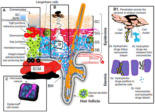
Table 1 Anti-inflammatory, antihypertensive, and anti-infective drugs incorporated in UDVs
Table 2 Other drugs incorporated in UDVs