
Figures & data
Figure 1. Magnetic resonance imaging of the spine.
(A) Post-contrast T1-weighted sagittal image of initial tumor. (B) T1 Dixon sagittal image of recurrent tumor (12 years later) with extramedullary extradural soft tissue mass between C7 and T1. (C) Post-contrast T1-weighted axial image of initial tumor. (D) STIR sagittal image of recurrent tumor.
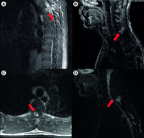
Table 1. Comprehensive overview of the patient's clinical course, detailing the initial diagnosis, treatment modalities employed at various stages and the response to therapies.
Figure 2. The patient's atypical teratoid rhabdoid tumor (AT/RT) on histology.
H&E sections demonstrate a malignant neoplasm characterized by cells with vesicular chromatin and visible nucleoli in both the initial resection and the re-resection (A, C). Immunohistochemistry for BAF47 (INI1) shows loss of expression in tumor cells in both the initial resection (B) and the re-resection (D). Immunohistochemistry performed on the re-resection was positive for vimentin (E), smooth muscle actin (F), and EMA (G). PDL-1 (22C3) performed on the re-resection was positive in approximately 30% of neoplastic cells (H).
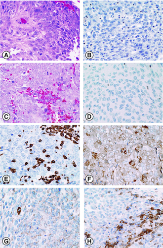
Table 2. Concise overview of the patient's differential diagnosis, covering considerations for both initial and recurrent tumors, immunohistochemistry findings and final diagnosis.