Figures & data
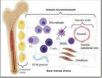
The bone marrow microenvironment is divided into endosteal, and vascular niches set within a stroma of differentiated accessory or ‘stromal’ cells, such as fibroblasts, osteoclasts, osteoblasts, adipocytes, endothelial cells, macrophages and mast cells as well as ECM proteins. As for the term ‘immune microenvironment’, it refers to a functional compartment of differentiated immune cells located throughout the marrow stroma.
DC: Dendritic cell; ECM: Extracellular matrix; MDSC: Myeloid-derived suppressor cell; PC: Plasma cell.
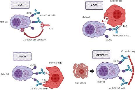
Top left: mAbs bind CD38. The Fc fragment is bound by C1q, initiating the complement cascade, and resulting in a membrane attack complex, leading to cell lysis and death. Top right: mAbs bind CD38. The Fc fragment is then bound by an FcR-bearing effector cell, such as a natural killer cell, leading to activation of cytotoxic processes. Bottom left: mAbs bind CD38, and its Fc fragment is then bound by an FcR-bearing macrophage, inducing phagocytosis. Bottom right: FcR-mediated crosslinking of mAbs induces direct cellular apoptosis.
ADCC: Antibody-dependent cell-mediated cytotoxicity; ADCP: Antibody-dependent cellular phagocytosis; CDC: Complement-dependent cytotoxicity; MAC: Membrane attack complex; MM: Multiple myeloma.