
Abstract
The sinuses of Valsalva are three pouch-like dilations at the root of the aorta, just above the aortic valve. Aneurysms of the sinuses of Valsalva can be congenital or acquired secondary to infective endocarditis. Here, we describe the case of a 41-year-old male patient who presented to the cardiology department of our hospital with fatigue and dyspnea on exertion for long periods. He had a history of infective endocarditis, smoking and substance abuse, like khat. Cardiovascular examination showed a murmur consistent with aortic regurgitation. Other systemic examinations, including those of the neurological, respiratory and abdominal, were unremarkable. Laboratory investigations were normal. Cardiac imaging, like transthoracic, trans-esophageal and cardiac computed tomography showed an aneurysm of the sinus Valsalva dissecting in to the interventricular septum. The patient was managed with metoprolol 25 mg, perindopril 10 mg and furosemide 20 mg and recommended for surgery but unfortunately it is not available in our country.
Introduction
The sinuses of Valsalva (SOV) are three pouch-like dilations at the root of the aorta, just above the aortic valve. These sinuses play a crucial role in the proper functioning of the heart and the aortic valve.Citation1 Aneurysms are weaknesses in the walls of the sinuses that can lead to the development of aneurysms. Aneurysms of the sinuses of Valsalva can be congenital or acquired, and they may be associated with conditions such as connective tissue disorders. It can potentially lead to complications such as aortic regurgitation or compression of nearby structures.Citation2,Citation3 These patients are usually asymptomatic if the aneurysm is intact. Those with ruptured or dissected aneurysms develop symptoms like chest pains or shortness of breath (SOB)Citation4 and surgical repair is usually mandatory in cases where aneurysms are encroaching on nearby structures and causing myocardial ischemia. Ruptured SOV aneurysms (RSOV) are rare, and their dissecting into the interventricular septum (DAIS) is scarcely reported.Citation5 Here we present a 40-year-old male with a sinus Valsalva aneurysm dissecting into the interventricular septum as a complication of infective endocarditis.
Case Presentation
A 41-year-old male with a history of infective endocarditis and social history of heavy smoking and khat chewing presented to a cardiology outpatient with fatigue and dyspnea on exertion.
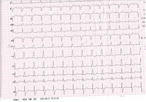
Vital signs at presentation were in the normal range, except for a heart rate of 110 bpm and mild grade fever (36.9 oC). Systemic review of neurology, respiratory, and abdominal were unremarkable. On cardiac examination, he had a diastolic murmur at the level of the aorta, consistent with aortic regurgitation. The laboratory tests of thyroid function, full blood count, renal function, and liver function were in the normal range. Complete right bundle branch block (RBBB) with left axis deviation (ALD) consistent with a bi-fascicular block was seen on electrocardiography ().
Figure 1 Electrocardiography showing bi-fascicular block.
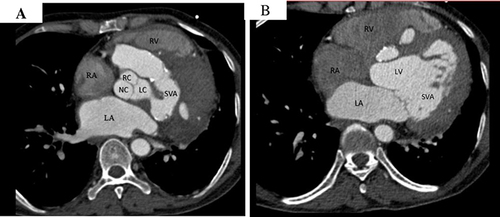
Echocardiography demonstrated a right sinus Valsalva aneurysm with multiple interventricular septum abscess like vegetation ( and ). Further cardiac CT assessment showed a larger sinus Valsalva aneurysm and marginal calcification with suspicious communications into the interventricular septum. ( and ).
Figure 2 (A) Parasternal short axis view sinus Valsalva aneurysm dissecting in to interventricular septum (red arrow), (B) Apical 4 chamber view interventricular aneurysm (red arrow).
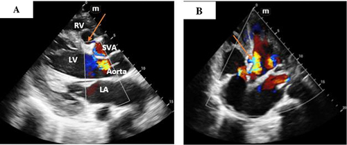
Figure 3 Cardiac CT in axial view (A and B) Showing left sinus of Valsalva aneurysm with suspicious communication in to interventricular septum.
The case was managed with metoprolol 25 mg, ramipril 2.5 mg 2×1, and furosemide 20 mg and recommended cardiac surgery but unfortunately it is not available in our country. Though he was advised to go where cardiothoracic surgery is available, the patient did not go due to financial constraints, and he is now on cardiology outpatient for his fellow up.
Discussion
A rare case of a right sinus Valsalva aneurysm dissecting into the interventricular septum due to infective endocarditis is a complex and unusual condition. This case involves the rupture of the sinus of Valsalva aneurysm, leading to complications such as interventricular septal dissection and infective endocarditis. The literature indicates that such cases are extremely rare and often associated with high mortality rates.Citation6 The sinus of Valsalva aneurysm is a congenital cardiac anomaly, but it can also be secondary to chest trauma, infection, aortic root involvement, syphilis, Behçet disease, atherosclerosis, and Marfan syndrome.Citation7 The rupture of the sinus of Valsalva aneurysm can lead to various complications, including right ventricular outflow tract obstruction, myocardial infarction due to coronary artery compression, conduction disturbances, and endocarditis.Citation8
Echocardiography and CT findings play a crucial role in diagnosing and understanding the extent of the sinus of Valsalva aneurysm and its complications. Three-dimensional echocardiography has been particularly useful in delineating the anatomy of ruptured sinus of Valsalva aneurysms, especially in cases of infective endocarditis.Citation9 Additionally, transesophageal echocardiography has been instrumental in the surgical management of pseudoaneurysms of the mitral-aortic intervalvular fibrosa with aneurysms of the right sinus of Valsalva and the left main coronary artery.Citation10
Comparing this case to other reported cases, it is evident that the involvement of the interventricular septum due to the dissecting right sinus Valsalva aneurysm is a rare and unique occurrence. While most reported cases focus on the rupture of the sinus of Valsalva aneurysm into the right atrium or ventricle, the involvement of the interventricular septum presents distinct challenges and considerations for surgical management and treatment. The rarity of this presentation underscores the importance of comprehensive imaging and diagnostic modalities to accurately assess the extent of the aneurysm and its complications.Citation11
Conclusion
The case of a sinus Valsalva aneurysm dissecting into the interventricular septum due to infective endocarditis is a rare and complex condition that requires multidisciplinary management involving cardiology, cardiac surgery, and imaging specialists. The unique nature of this case highlights the need for further research and comprehensive reporting of similar cases to enhance our understanding and management of this rare cardiac anomaly.
Consent
Written informed consent was obtained from the patient’s family to have the case details and any accompanying images published.
Disclosure
The authors report no conflicts of interest in this work.
Additional information
Funding
References
- Xu B, Kocyigit D, Betancor J, et al. Sinus of valsalva aneurysms: a state-of-the-art imaging review. J Am Soc Echocardiography. 2020;33(3):295–312. doi:10.1016/j.echo.2019.11.008
- Nguyen Q, Vervoort D, Phan K, et al. Surgical management for unruptured sinus of valsalva aneurysms: a narrative review of the literature. J Thoracic Dis. 2021;13(3):1833–1850. doi:10.21037/jtd-20-2682
- Nunes SV, Arruda AD, Rodrigues YL, et al. Giant ruptured sinus of valsalva aneurysm. In: Cardiovascular Surgery: A Clinical Casebook. Springer;2019:37–45.
- Wu P, Yu S, Zeng J, et al. Aortic sinus aneurysm invading ventricular septum and dissection caused by Behcet’s disease: a case report and literature review. BMC Cardiovasc Disord. 2023;23(1):429. doi:10.1186/s12872-023-03420-7
- Ghosh S, Bootla D, Barward P, et al. Multilobulated sinus of valsalva aneurysm Dissecting into the Interventricular Septum (DAIS) and rupturing into left ventricle: a case report. Eur Heart J Case Rep. 2022;6(2):ytac019. doi:10.1093/ehjcr/ytac019
- Abetti A, Gandet T, Amri A, et al. Ruptured right valsalva sinus into the right atrium due to infective endocarditis: a case report. Pan Afr Med J. 2020;37. doi:10.11604/pamj.2020.37.65.21491
- Barroso F, Takakura I, Reis R, et al. Ruptured aneurysm of the sinus of valsalva causing pulmonary embolism: a rare association. Aorta. 2020;08(04):107–110. doi:10.1055/s-0040-1722099
- Chandra S, Vijay S, Dwivedi S, Saran R. Delineation of anatomy of the ruptured sinus of valsalva with three-dimensional echocardiography: the advantage of the added dimension. Echocardiography. 2012;29(6):E148–E151. doi:10.1111/j.1540-8175.2011.01652.x
- Güler N, Eryonucu B, Tuncer M, Aşker M. Aneurysm of sinus of valsalva dissecting into interventricular septum: a late complication of aortic valve replacement. Echocardiography. 2004;21(7):645–648. doi:10.1111/j.0742-2822.2004.03128.x
- Haque H, Afroz F, Afroze S, et al. Ruptured sinus of valsalva aneurysm complicating infective endocarditis: a case report. Birdem Med J. 2016:52–55. doi:10.3329/birdem.v5i3.28540
- Joshi S, Jagadeesh A, Furtado A, Bhat S. Transesophageal echocardiography in surgical management of pseudoaneurysm of mitral-aortic intervalvular fibrosa with aneurysms of right sinus of valsalva and left main coronary artery. Ann Card Anaesth. 2013;16(1):40. doi:10.4103/0971-9784.105368