
Figures & data
Figure 1. Schematic presentation of small intestine. Particles may enter the intestinal barrier from the small intestine lumen through microfold cells (M-cells) that present them to immunological cells (i.e. dendritic cells) in the lamina propria and the Peyer’s patches. Peyer’s patches are rich in T cells, macrophages, and activated antibody-secreting B and plasma cells. The single layer of the intestinal epithelium is protected by mucus produced by Goblett cells and containing mucin glycoproteins where immunoglobulin A (IgA) and antimicrobial peptides prevent interaction of microbiota with the cell surface. Disruption of the intestinal barrier leads to the secretion of pro-inflammatory cytokines by T-cells (adopted from [Citation29]).
![Figure 1. Schematic presentation of small intestine. Particles may enter the intestinal barrier from the small intestine lumen through microfold cells (M-cells) that present them to immunological cells (i.e. dendritic cells) in the lamina propria and the Peyer’s patches. Peyer’s patches are rich in T cells, macrophages, and activated antibody-secreting B and plasma cells. The single layer of the intestinal epithelium is protected by mucus produced by Goblett cells and containing mucin glycoproteins where immunoglobulin A (IgA) and antimicrobial peptides prevent interaction of microbiota with the cell surface. Disruption of the intestinal barrier leads to the secretion of pro-inflammatory cytokines by T-cells (adopted from [Citation29]).](/cms/asset/03183c34-a441-40ff-a6cf-0e55fd9c855e/yrer_a_2333619_f0001_oc.jpg)
Figure 2. Schematic of the intestinal crypt-villus unit. The intestinal crypt-villus unit is maintained by multipotent crypt base columnar (CBC; Lgr5 +) and +4 cells (Hopx +, Bmi1 +, mTert +, Lrig +). These stem cells are found in the crypt and supply the villus with specialized intestinal cells, including enterocytes, goblet cells, enteroendocrine cells (EEC), and tuft cells, which are eventually shed at the villus tip. Conversely, Paneth cells are mature cells that remain in the crypt and modulate the stem cell environment. Figure adapted, with permission from Company of Biologists, from Beumer et al. 192. Available herein via license: Creative Commons Attribution 4.0 International from https://www.researchgate.net/publication/323367542_Comparative_regenerative_mechanisms_across_different_mammalian_tissues.
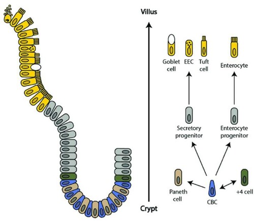
Figure 3. Schematic of tight junctions in the healthy gut. Tight junctions are at the apical end of junctional complexes composed of three transmembrane proteins: occludin claudins and junctional adhesion molecules (JAMs) that bind intracellular membrane proteins, zonula occludens (ZOs) which connect the transmembrane tight junction to the actin skeleton. Below the tight junctions are adherens junctions composed of transmembrane proteins, E-cadherin and nectin linked to the cytoskeleton by scaffolding proteins, catenin and afadin linked to actin filaments. Desmosomes are made up of the transmembrane liner glycoproteins, desmoglein and democollin which are cadherin proteins linked to intermediate keratin filaments. Gap junctions form a tunnel for small molecules to pass between adjacent epithelial cells. Image made using BioRender. Available via license: Creative Commons Attribution 4.0 International from https://www.researchgate.net/publication/348420396_Role_of_Metabolic_Endotoxemia_in_Systemic_Inflammation_and_Potential_Interventions.
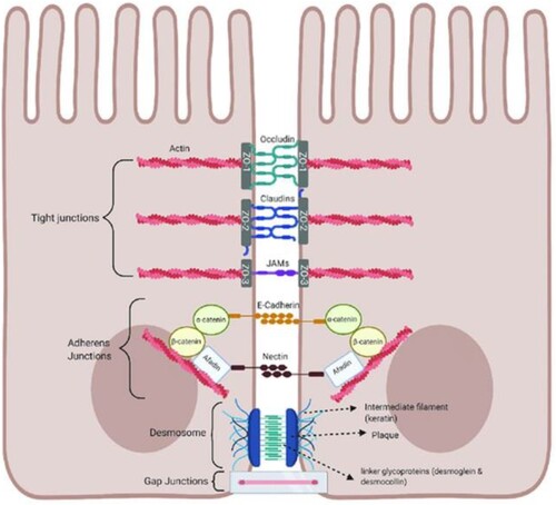
Figure 4. Nanosystems (nanocarriers) studied in nanomedicine and corresponding applications in healthcare.
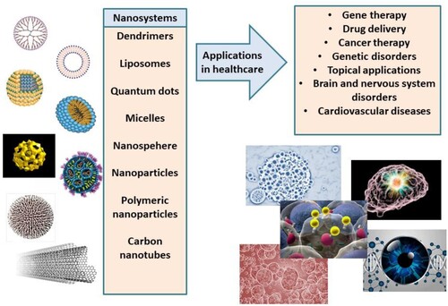
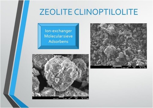
Figure 5. Electron micrographs of zeolite particles that can serve as ion exchangers, molecular sieves as well as an adsorbent. Images were obtained by SEM Jeol JSM-7800F.
Data availability statement
All data and references used for the preparation of the manuscript have been derived from public domain resources.