
Figures & data
Table 1 Rewards provided by myrmecophytic plants
Figure 1 Rewards produced by myrmecophytic plants to establish stable obligate symbiotic mutualisms with ants.
Notes: Extrafloral nectar of Acacia hindsii (A) and Acacia cornigera (B). Food bodies and a hollow thorn of Acacia sp. (C). A hollow stem of Macaranga bancana (D). Food bodies at the base of a leafstalk in Cecropia mexicana (E).
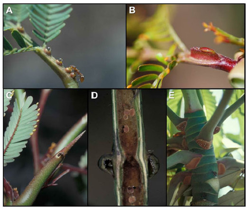
Table 2 Plant–ant obligate interactions in six plant groups
Figure 2 Anatomical tissues typically recognized in extrafloral nectaries.
Abbreviations: E, epidermis; NP, nectary parenchyma; SNP, subnectary parenchyma.
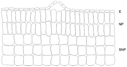
Figure 3 Comparison of the anatomical structure of food bodies in Piper and Acacia cornigera.
Notes: In Piper (A), FBs are anatomically simple and involve the enlargement of single epidermal cells without any involvement of the underlying parenchyma. In Acacia cornigera (B), FBs are anatomically differentiated and show a specialized internal differentiation of cells. They are connected to the vascular tissue.
Abbreviations: EC, epidermal cell; E, epidermis; CC, cortex cell; FBs, food bodies; H, hypodermis, VS, vascular system.
Abbreviations: EC, epidermal cell; E, epidermis; CC, cortex cell; FBs, food bodies; H, hypodermis, VS, vascular system.
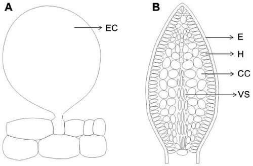
Figure 4 Comparison of the anatomical structure between a hallow and an unhollowed stem.
Notes: The figure depicts differences in the anatomical structure between a hollow stem of a myrmecophytic plant (A) and an unhollowed stem of a myrmecophilic plant (B). Copyright © 2007 The Botanical Society of America. Adapted from Tepe EJ, Vincent MA, Watson LE. Stem diversity, cauline domatia, and the evolution of ant–plant associations in Piper sect. Macrostachys (Piperaceae). Am J Bot. 2007;94:1–11.Citation98
Abbreviations: C, crystal; VB, vascular bundle; WT, wound tissue.
Abbreviations: C, crystal; VB, vascular bundle; WT, wound tissue.
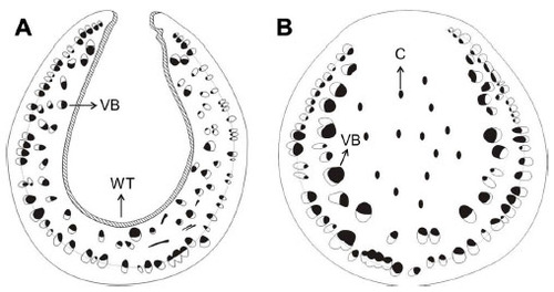
Figure 5 Comparison of the anatomical structure of an ant domatia with that of a leaf lamina.
Notes: The figure depicts differences in the anatomical structure between an ant domatia (A) and a leaf lamina (B).
Abbreviations: PP, palisade parenchyma; E, epidermis; PT, parenchymatous tissue; PP, palisade parenchyma; SP, spongy parenchyma; VB, vascular bundle.
Abbreviations: PP, palisade parenchyma; E, epidermis; PT, parenchymatous tissue; PP, palisade parenchyma; SP, spongy parenchyma; VB, vascular bundle.
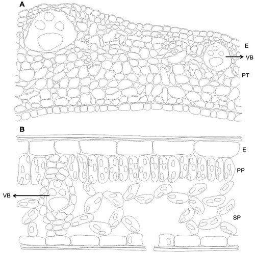
Table 3 Outstanding questions