

Abstract
EWSR1 is a member of the FET family of nucleic acid binding proteins that includes FUS and TAF15. Here, we report the systematic analysis of endogenous EWSR1’s cellular organization in human cells. We demonstrate that EWSR1, which contains low complexity and nucleic acid binding domains, is present in cells in faster and slower-recovering fractions, indicative of a protein undergoing both rapid exchange and longer-term interactions. The employment of complementary high-resolution imaging approaches shows EWSR1 exists in two visual modalities, a distributed state which is present throughout the nucleoplasm, and a concentrated state consistent with the formation of foci. Both EWSR1 visual modalities localize with nascent RNA. EWSR1 foci concentrate in regions of euchromatin, adjacent to protein markers of transcriptional activation, and significantly colocalize with phosphorylated RNA polymerase II. Our results contribute to bridging the gap between our understanding of the biophysical and biochemical properties of FET proteins, including EWSR1, their functions as transcriptional regulators, and the participation of these proteins in tumorigenesis and neurodegenerative disease.
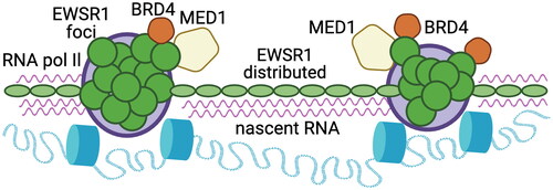
Graphical Abstract
Acknowledgments
We are grateful to Javed Khan (Genetics Branch), and members of the Caplen laboratory for discussions and comments on the manuscript. We also thank Sanjit Mukherjee and Roshan Shrestha (Genetics Branch) for their technical advice and guidance and the CCR Sequencing Facility (long-read sequencing, Pac Bio), CCR Genomics Core (Sanger sequencing), and CCR Flow Cytometry Core, especially Karen Wolcott, for technical assistance. The content of this publication does not necessarily reflect the views or policies of the US Department of Health and Human Services, nor does mention of trade names, commercial products, or organizations imply endorsement by the US Government.
Author contributions
S.S. Rajan and N. J. Caplen conceived and designed this study. S. S. Rajan conducted most experiments and analyzed the data. J. Loncarek supported the employment of microscopy (SIM and STED), performing experimentation and image and data analysis. V. Ebegboni performed the IP experiments, P. Pichling generated immunoblotting results, and K. Ludwig assisted with generating and characterizing the modified EWS cell lines. T. L. Jones contributed to the long-read sequence analysis of the modified EWS cell lines. R. Chari designed and generated the CRISPR-targeting reagents. M. Kruhlak contributed technical support and advice, particularly the employment of confocal and super-resolution microscopy, including FRAP analysis. A. Tran assisted with image analysis. N. J. Caplen supervised the project and conducted some of the data analysis. S. S. Rajan and N. J. Caplen wrote the manuscript. All the authors read and agreed on the publication of this manuscript.
Disclosure statement
No potential conflict of interest was reported by the authors.
Data availability statement
The gene-specific long-read sequencing from this study has been deposited to the DRYAD database (https://datadryad.org) and assigned the identifier doi:10.5061/dryad.fqz612jz3.