Figures & data
Figure 1. A CT scan of chest and abdomen on the day of admission. A mild inflammation was seen in the lower lobes of bilateral lungs, and the liver was slightly enlarged.
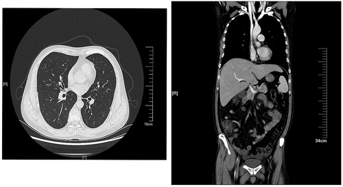
Figure 2. Histoplasma capsulatum (numerous, capsulated yeast cells) of phagocyte cells (arrow) in bone marrow smear (Wright’s staining, magnification 1,000×).
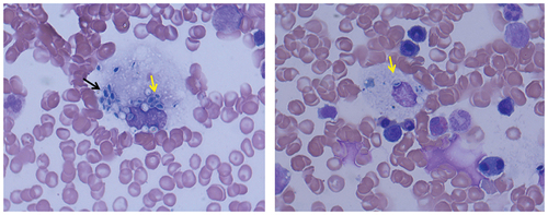
Figure 3. (a, b) Yeast-like spores and hyphae (with an arrow, gram staining, magnification 1,000×) under microscopy with bone marrow cultured at 37 °C in 16 days. (c, d) Colonies grown at 28 °C and 37 °C with bone marrow bone cultured in 16 days; (c) White, short villous and filamentous colonies; (d) Yeast-like colonies. (e, f) Fungal morphology under microscopy by taking colonies, respectively; (e) A filamentous form with tuberculate macroconidia on the septate hyphae (lactic acid phenol cotton blue staining); (f) Yeast-like fungus, budding cells with pseudohyphae (gram staining).
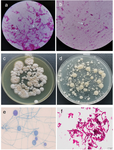
Table 1. The regional distribution of 225 patients diagnosed as histoplasmosis from 2012 to 2022 in China.
Table 2. The clinical characteristics of 225 patients diagnosed as histoplasmosis from 2012 to 2022 in China.
Table 3. The diagnosis, treatment, and outcome of 225 patients diagnosed as histoplasmosis from 2012 to 2022 in China.
Figure 4. Modified PRISMA flow chart.
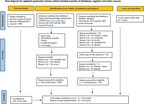
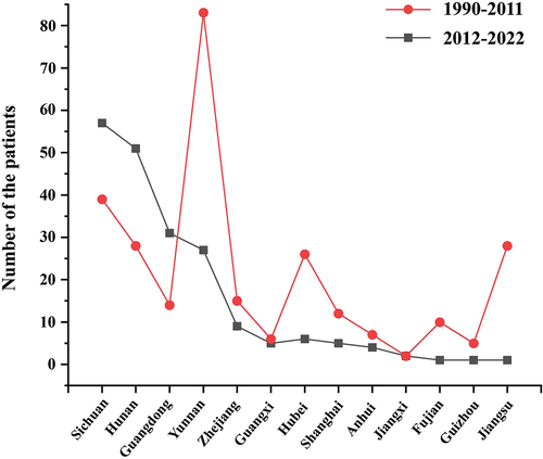
Figure 5. The geographical distribution of histoplasmosis diagnosed in southern China during 2012–2022 compared to those during 1990–2011.