Figures & data
Figure 1 Orthopedic implants, such as the total knee replacement components shown left, have sufficient size and volume for placement of sensors (S), electronics (E) and antenna (A) components within.
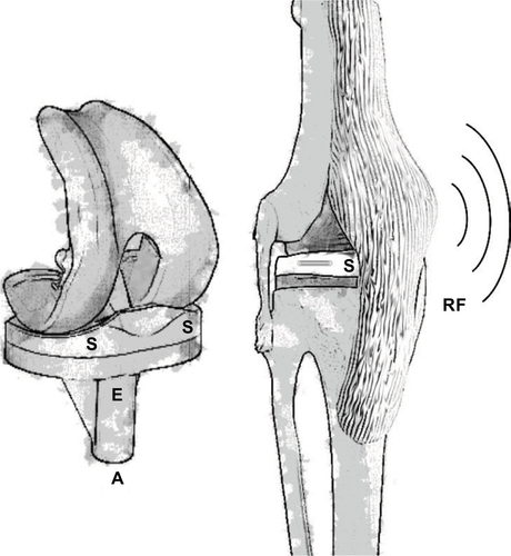
Figure 2 Strain gages are mounted onto the surface of implants such as the cervical spinal interbody cage shown and require lead wires connecting them to signal conditioning electronics.
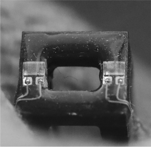
Figure 3 The technology that enables smart orthopedic implants has evolved over several decades, as shown in the total hip prostheses, from tethered electronics (left) to wireless systems that require signal conditioning electronics (E) with antennas (A) to be housed inside the implant (center) to passive sensors (P) that require no electronics and little-to-no modification of the implant (right).
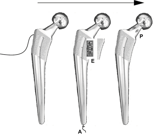
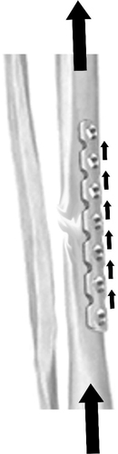
Figure 4 When a fracture is treated with open reduction and internal fixation, forces applied through the bone are transmitted through the plate.