Figures & data
Figure 1 Digestive tract of Apis mellifera adult worker.
Notes: (A) Anatomical view of digestive tract showing insertion of Malpighian tubules (arrow) between the mg and the hg. (B) Details of Malpighian tubules (arrows) (Photos courtesy of Pamela Decio).
Abbreviations: R, rectum; mg, midgut; hg, hindgut; P, proventriculus.
Abbreviations: R, rectum; mg, midgut; hg, hindgut; P, proventriculus.
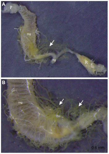
Figure 2 MT of Apis mellifera larvae; Histological sections stained with hematoxylin–eosin.
Notes: (A) End of the larval feeding phase showing the MTs connected to transition between the mg and the pg. (B) Distal portion of the MTs with flattened cells and larger lu containing excretory products. (C) Proximal portion of MTs during the end of larval postdefecation phase showing vacuolization of the epithelium of the tubule.
Abbreviations: MT, malpighian tubule; mg, midgut; pg, posterior gut; lu, lumen; tr, trophocyte of fat body; en, oenocyte of fat body.
Abbreviations: MT, malpighian tubule; mg, midgut; pg, posterior gut; lu, lumen; tr, trophocyte of fat body; en, oenocyte of fat body.
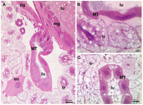
Figure 3 MTs of adult worker Apis mellifera; Histological sections stained with Hematoxylin-Eosin.
Notes: (A) MTs connected between the mg and pg. (B) Details of MTs showing epithelium with nu cells surrounding the lu. (C) Whole mount preparation of MT of Melipona scutellaris (Hymenoptera, Apidae) worker. Confocal images with faloidin showing F-actin in green (Cy5) and nuclei in cyan (DAPI).
Abbreviations: MT, Malpighian tubule; mg, midgut; pg, posterior gut; nu, mononucleated; lu, lumen.
Abbreviations: MT, Malpighian tubule; mg, midgut; pg, posterior gut; nu, mononucleated; lu, lumen.
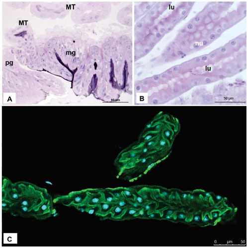
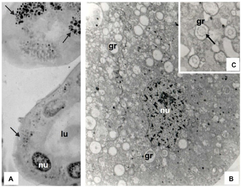
Figure 4 MT of an adult worker Atta sexdens rubropilosa.
Notes: (A) Histological sections stained with hematoxylin–eosin. Note the granules (black arrows) spread by the cytoplasm of excretory cells. (B and C) Transmission electron microscopy. General view of excretory cell in (B) highlighting the gr and nu. Detail of granules in (C) showing the concentric ring (black arrow) inside them.
Abbreviations: MT, Malpighian tubule; nu, nucleus; lu, lumen; gr, granules.
Abbreviations: MT, Malpighian tubule; nu, nucleus; lu, lumen; gr, granules.