
ABSTRACT
The quest for natural and sustainable ingredients in cosmetics has led to increased interest in marine-derived biomaterials for skin rejuvenation. The marine ecosystem holds a plethora of unique organisms that produce bioactive compounds with potential benefits for human skin health and rejuvenation. This manuscript examines the diverse potential of marine collagen, sourced from jellyfish, sponge collagen (spongin), and fish collagen, as a key component in skin rejuvenation. Jellyfish collagen demonstrates moisturizing effects and protection against UV-induced photo aging, while sponge collagen exhibits promising applications in promoting cell proliferation and mitigating inflammatory responses. Fish collagen, particularly from fish skin sourced from processing waste, stands out for its safety, moisture retention, and protective effects against UV radiation. This review underscores the significance of marine collagen in enhancing skin hydration, renewal, UV protection, cell proliferation, and inflammation reduction. With extensive clinical evidence supporting these positive effects, the cosmetic industry is poised to leverage the regenerative capabilities of marine collagen for the development of safe, effective, and holistic skin rejuvenation solutions.
Introduction
Cosmetics are substances made up of mixtures of chemicals obtained from either natural sources or created synthetically for external usage in various parts of the body for cleaning, perfuming or altering their appearance [Citation1]. They serve a variety of purposes, including cleansing the body, protecting the skin, concealing blemishes, enhancing a person’s natural beauty, and changing the entire appearance of a person [Citation2]. Early users of cosmetics include Egyptians, who use kohl and castor oil for skin protection, and Romans, who use beeswax, olive oil, and rosewater for the same purpose, long before the advent of vaseline and lanolin in the 19th century [Citation3].
The cosmetic industry has been proliferating at an alarming rate in the 21st century [Citation2], generating an estimated global market volume of over 70 billion dollars in 2001 and expected to rise beyond 400 billion dollars by 2028 [Citation4,Citation5]. This rapid increase has been associated with social influence and the desire for social acceptance [Citation6]. Traditional cosmetic products are expected to exhibit appropriate dermatological efficacy while maintaining a high level of safety during use [Citation7]. However, synthetic cosmetic formulations, on the other hand, may have the intended results, they are often associated with side effects such as hypo-or hyperpigmentation, infection, erythema and solar lentigines [Citation8–10]; nonetheless, empirical studies have revealed the incidence of noteworthy adverse responses within this group. For example, hydroquinone, a commonly used skin-lightening chemical, has been shown to have mutagenic properties and the ability to produce ochronosis [Citation6]. Furthermore, several components such as kojic acid, which is well known for its role in skin thickening, have been associated with carcinogenic qualities [Citation2]. Notable examples include p-phenylenediamine, a component of commercial dyes linked to blistering and edema, and butylated hydroxyanisole, a component found in lipsticks and moisturizers that have been classified as carcinogenic by the International Agency for Research on Cancer [Citation6]. Consequently, the use of synthetic cosmetics is discouraged, encouraging the development of alternatives based on natural product-based cosmetic formulations. It has also been reported that natural product cosmetics are gaining increasing public attention due to the generally held belief that they are safer than synthetic cosmetics [Citation11].
In contemporary society, the quest for effective, efficient and safe skin rejuvenation procedures has become increasingly important, where a young look is generally connected with vigor and well-being. Skin aging is a complex physiological process influenced by intrinsic genetic factors and extrinsic environmental factors and manifests through the gradual loss of elasticity, firmness, and moisture. The skin frequently serves as a reflection of overall health and internal physiological status. Various internal disorders have the potential to impact the properties of the skin. Over time, skin alterations can influence self-esteem, quality of life, and social interactions [Citation12]. Consequently, the cosmetic and dermatological industries have seen an increased demand for creative ways to combat these indicators of aging and restore the youthful skin characteristics. One fascinating area that has gained traction is the use of biomaterials for anti-aging interventions.
Biomaterials have become essential in anti-aging techniques, because of their ability to provide structural support, preserve moisture, and improve chemical penetration in skin rejuvenation [Citation13,Citation14]. These compounds, which are frequently derived from natural sources, stimulate collagen formation, provide antioxidant protection, and integrate well with skin tissues, thereby decreasing immunogenicity and toxicity concerns [Citation15,Citation16].
For skin rejuvenation, a wide range of biomaterials are used to address various aging issues and improve skin appearance. These biomaterials include hyaluronic acid for moisture retention and volume restoration and collagen for improved elasticity and reduced wrinkles [Citation17,Citation18]. Marine ecosystems harbor a rich diversity of organisms that have evolved unique bioactive (biomaterial) compounds as adaptions for their underwater environments.
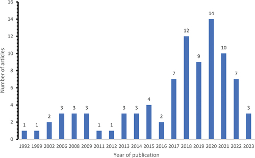
The use of marine-derived biomaterials in cosmeceuticals is an exciting new area in skincare, providing a multitude of natural compounds with enormous potential for improving skin health and appearance. From seaweed to specific fish species, the ocean is abundant with bioactives that demonstrate a promising avenue for tissue engineering, wound healing, and cosmeceutical products [Citation19]. Marine-derived compounds provide several advantages, including deep hydration, collagen stimulation, environmental protection, and even speeding up the skin’s healing process [Citation20]. Furthermore, their sustainable origin increases their attractiveness as ecologically friendly alternatives to traditional skincare ingredients. As research continues to explore the therapeutic effects of marine-derived cosmeceuticals (), the potential for leveraging the ocean’s potency in skin rejuvenation and care holds enormous promise for altering how we care for our skin. This review aims to explore the current scientific literature on marine-derived biomaterials for skin rejuvenation in cosmetics.
Figure 1. Number of articles published on the therapeutic effects of marine-derived cosmeceuticals.
Methodology
The literature research for this review incorporated published journal articles and books. Studies were identified by searching electronic databases such as Google Scholar, PubMed, ScienceDirect, Sci-Finder, and Scopus. Additionally, the reference lists of identified papers were reviewed to verify that the literature was thoroughly covered. Collectively, a total of 138 articles were included for this review and findings are presented narratively. For this review, only papers in English were evaluated.
Skin aging
As individuals age, the skin undergoes a complex series of cellular changes that manifest in various visible signs of aging. While preserving the skin’s structural and functional integrity is essential, certain crucial skin functions, such as defense mechanisms and healing capacity, decline over time [Citation21,Citation22]. Existing literature attributes this phenomenon to the progressive deterioration of skin physiology [Citation23]. Aged skin is characterized by diminished suppleness and fat content, alongside a reduction in collagen and elastin levels, resulting in a thinner and smoother appearance [Citation24]. Consequently, skin aging can have a detrimental impact on individuals’ self-confidence, potentially compromising their overall quality of life and social well-being [Citation25,Citation26]. As a result, addressing skin aging has become a top priority within the cosmetic and skincare industries.
Significant changes occur in fibroblasts, collagen, and elastic fibers in the skin aging process. Reactive oxygen species (ROS) generated in the aging process activate mitogen-activated protein kinases (MAPKs) and induce transcription factors, including activator protein 1 (AP-1) and nuclear factor-κB (NF-κB) [Citation27]. This activation increases matrix metalloproteinase (MMP) expression and inhibits transforming growth factor-β (TGF-β) signaling, which leads to collagen fragmentation and decreased collagen biosynthesis [Citation28]. This hinders the mechanical interaction between fibroblasts and the extracellular matrix (ECM) and consequently reduces the size of dermal fibroblasts. Aged fibroblasts produce a greater amount of ROS that further increases the expression of MMPs and inhibits TGF-β signaling, creating a positive feedback loop that accelerates dermal aging [Citation28]. MMP-12 secreted from fibroblasts and macrophages plays a crucial role in the development of solar elastosis and in the reduction of functional elastic fibers () [Citation29].
Figure 2. Schematic illustration showing the changes in the fibroblasts, collagen, and elastic fibers in skin ageing (adopted from [Citation29]).
![Figure 2. Schematic illustration showing the changes in the fibroblasts, collagen, and elastic fibers in skin ageing (adopted from [Citation29]).](/cms/asset/573b1955-a1ff-4619-9b45-a7b8008bdaf1/teba_a_2336307_f0002_oc.jpg)
highlights some of the intricate and interrelated cellular changes that occur in the skin. These changes occur in the deeper layers of the skin and are affected by variables such as genetics, environmental exposures, lifestyle choices, and general health. Whilst figure one illustrates the changes in fibroblasts, collagen, and elastic fibers in the dermal aging process.
Table 1. Clinical manifestation of skin aging.
Marine-derived biomaterials
Natural products encompass materials derived from plants, animals, and marine sources. Marine biomaterials are mainly derived from aquatic animals (fish, crustaceans, and mollusks), seaweeds, and sponges [Citation14,Citation38]. Biopolymers derived from these sources have been used in many biological and biomedical processes, including the cosmetic industry. Although the majority of natural cosmetic products are produced from plant resources [Citation39]; marine biomaterials are preferred in many industries, such as biomedical, tissue engineering and pharmaceutical [Citation14]. These are attributable to their applicable, accessible, and cost-effective benefits [Citation2]. Hence, marine biomaterials are now being researched globally as a promising source of cosmetics for skin rejuvenation because of the vast biological activities of extracts related to skin improvement [Citation40].
Biomaterials from marine sources have been found to elicit anti-inflammatory, antioxidant, and anti-pigmentation properties, making them valuable ingredients in the cosmetic industry for skin rejuvenation [Citation14,Citation17,Citation41]. Marine ecosystems account for about 70% of biosphere biodiversity, making them an excellent source of natural products [Citation42]. Among various natural organisms in the marine habitat, algae are the most abundant, with rapid growth and reproduction compared to terrestrial plants or higher plants. Therefore, they are valuable sources of bioproducts and metabolites for skin rejuvenation and health [Citation14,Citation16]. Other marine organisms, such as bacteria, fish, and poriferans (jellyfish, sponges), are repositories of a significant amount of collagen, an essential biomaterial for skin rejuvenation and health. Fish and poriferans are more desirable than other sources of marine biomaterials because they are metabolically compatible without constraints and are pathogen-free [Citation17].
When comparing the properties of marine cosmeceuticals with traditional skincare ingredients, it becomes evident that marine-derived biomaterials offer unique advantages. Marine-derived cosmeceuticals often contain bioactive compounds such as peptides, antioxidants, and polysaccharides [Citation43,Citation44] that can provide various skin benefits, including hydration, anti-aging [Citation45], wound healing [Citation46,Citation47] and UV protection [Citation47,Citation48]. These compounds may have higher bioavailability and potency compared to traditional skincare ingredients [Citation44]. According to Zhang, Kim [Citation49] due to the lack of biological cues in the case of synthetic materials, bioactive from natural sources have displayed superior biofunctionality over synthetic, as well as low immune response and excellent biocompatibility in biomedical applications. For instance, some synthetic ingredients such as tyrosinase inhibitors have shown restrictions due to concerns regarding their toxicity, limited stability, inadequate skin penetration, and reduced efficacy [Citation50]. Additionally, surfactants and antioxidants from synthetic material have shown to have negative effects on human health due to their toxicity and skin irritation, whilst marine-derived ingredients are biocompatible and have less low irritancy to the skin [Citation43]. When compared to traditions skincare ingredients, natural products provide significant chemical diversity, enhanced specificity, and binding efficiency, making them attractive candidates for cosmetic applications [Citation51].
Fish oil for skin rejuvenation
Marine fish is a known source of oils; fish oils found relevance in the cosmetic industry due to polyunsaturated fatty acids content, such as omega-3 polyunsaturated fatty acids, docosahexaenoic acid (DHA), and eicosapentaenoic acid (EPA)of the oils which enhance and promote skin health and rejuvenation [Citation52]. The oil can be extracted from the frame, head, hepatic region, liver trimming, viscera, and skin of fish [Citation53]. The oil extracted from the hepatic region is the most desirable for cosmetological purposes because it is high in lipids, therefore most fish oils are extracted from the fish liver [Citation52].
Oral administration of fish oil supplements has been reported to help in skin rejuvenation and care. In a clinical study conducted by Khayef, Young [Citation54], they found that fish supplements reduce the inflammation of acne by inhibiting cytokine production in the body. A diet rich in fish oil lipid source was reported to increase the minimum erythema dose in patients with skin reactions triggered by infection or medicine Orengo, Black [Citation55]. Liver and skin oil rich in omega-3 from the marbled rock cod fish improved skin health and rejuvenation by inhibiting matrix metalloproteinase-1 (MMP-1) formation in an in vivo study conducted by Lee, Koo [Citation56].
Photoaging, a skin defect induced by exposure to ultraviolet (UV) rays from sunlight, can lead to adverse effects such as sunburn, photosensitivity, and inflammation. The Omega-3 polyunsaturated fatty acids (PUFAs) found in fish oil have been identified as a potential mitigator of these effects. The major PUFAs in fish oil are comprised of linoleic acid (LA), α-linolenic acid (ALA), DHA, and EPA [Citation52]. They work by inhibiting the production of pro-inflammatory eicosanoids, engaging in direct competition with arachidonic acid (AA) metabolism, as suggested by studies [Citation52,Citation57]. Moreover, the Omega-3 PUFAs play a role in decreasing the production of pro-inflammatory eicosanoids by competitively interacting with AA metabolism [Citation57]. These fatty acids exhibit various mechanisms in suppressing UV-induced keratinocyte damage, including the regulation of pathways such as cyclooxygenase-2 (COX-2), nuclear factor kappa B (NF-Κb), and mitogen-activated protein kinase (MAPK)/extracellular-signal-regulated kinase (ERK) [Citation58]. Through these mechanisms, Omega-3 PUFAs from fish oil demonstrate a potential protective effect against the adverse impacts of UV exposure on the skin [Citation59].
With regards to hyperpigmentation, skin hyperpigmentation by melanogenesis is stimulated by UV exposure, endothelin-1, α-melanocyte-stimulating hormone (α-MSH), growth factors, and cytokines [Citation60]. DHA has been found to decrease α-MSH-activated melanin production and increase tyrosinase degradation without affecting Microphthalmia-associated transcription factor (MITF) expression however has no influence on cell viability [Citation61]. ALA and LA are reported to reveal skin-whitening capability through the mechanism of tyrosinase inhibition [Citation52].
Apart from photoaging, fish oil plays a significant role in the prevention of cutaneous carcinogenesis or skin cancer, dermatitis, and skin wounds [Citation62]. Fish oils are helpful in the inhibition of skin cancer. For example, fish oil rich in n-3 fatty acids was found to inhibit papilloma formation at the promotion phase of skin cancer in mice [Citation62]. Fish oil and the related fatty acids are reported to be useful for ameliorating dermatitis symptoms. Barcelos, de Mello-Sampayo [Citation63] reported the ameliorative effect of fish oil rich in fatty acid on dermatitis symptoms, a decrease in cutaneous dryness and pruritus was observed after the oral administration of fish oil as supplementation in rats.
With regard to wound healing, the appropriate inflammation in the wound area promotes cell migration and skin tissue repair. The inflammatory phase of wound healing involves intricate cellular and molecular processes that are significantly influenced by proinflammatory cytokines. Notably, the synthesis and activity of these cytokines can be regulated by polyunsaturated fatty acids (PUFAs) [Citation64]. Omega-3 and omega-6, have been demonstrated to play a crucial role in shaping cell membrane structure and facilitating anabolic events essential for the reconstruction of skin tissue [Citation64]. The presence of these fatty acids suggests the possibility that they modulate or enhance the local inflammatory response at wound sites, potentially contributing to an accelerated rate of healing [Citation65]. The solid emulsion gel for cell-targeted poly-unsaturated fatty acid delivery to skin wounds described by Shingel, Faure [Citation66], showed a faster wound closure compared to the gel containing olive oil in an in vivo study using pigs.
Fish oil with its active components like omega-3 and omega-6 polyunsaturated fatty acids (PUFAs), has demonstrated efficacy in maintaining skin homeostasis and addressing various cutaneous issues. The fatty acids present in fish oil contribute to enhanced skin barrier function, inhibition of UV-induced inflammation and hyperpigmentation, alleviation of dry skin and pruritus associated with dermatitis, acceleration of skin wound healing, and prevention of skin cancer development [Citation52]. Importantly, these benefits can be realized through diverse administration routes, encompassing oral supplementation and topical application. However Huang, Wang [Citation52] highlights that the abundance of the fish genus complicates quality control, the precise fish type and the PUFA content in the fish oil are crucial aspects to consider for the skin advantages. Another factor to consider is that not only PUFAs but also vitamin A, vitamin D, retinol, selenium, and other components may contribute to fish oil’s bioactivity [Citation67]. Also, the most often expressed concern with omega-3 PUFA oral administration is the possibility of increasing the risk of bleeding due to the anti-platelet impact. In certain situations, dietary fish oil has been linked to gastrointestinal problems [Citation52].
Algae and seaweed extracts for skin rejuvenation
Algae and seaweed extracts have emerged as valuable resources in cosmetic science, particularly in the pursuit of effective skin rejuvenation. These natural wonders, hailing from aquatic environments, hold a rich reservoir of bioactive compounds that have shown immense promise in enhancing skin health and combating signs of aging [Citation68].
Algae, similar plants, thrive through photosynthesis and housed chloroplasts. The taxonomic landscape reveals a staggering array of approximately 20,000 algal species, meticulously enumerated and categorized [Citation69,Citation70]. Marine algae can be classified based on their size, giving rise to microalgae and macroalgae, which are commonly known as seaweeds. Microalgae are unicellular organisms; however, macroalgae have multicellular structures that are simpler than those observed in terrestrial plants [Citation71,Citation72]. Pigment analysis separates marine macroalgae into three different groups: Chlorophyta (green macroalgae with chlorophyll b and xanthophylls), Phaeophyceae (brown macroalgae with chlorophyll c and fucoxanthin), and Rhodophyta (red macroalgae with chlorophyll d and phycoerythrin) are the three major groups of algae [Citation70,Citation73]. Marine algae contain a diverse array of biomolecules, including amino acids, lipids, proteins, flavonoids, and carbohydrates [Citation74,Citation75].
The wealth of marine carbohydrates, coupled with their bioactivities, has ushered them into the spotlight of the cosmeceutical industry, particularly for skin treatments [Citation76]. Notably, the marine algae carbohydrates are fucoidan, laminaran, porphyran, and carrageenan, each offering distinct anti-aging and skin-enhancing effects [Citation69,Citation77,Citation78].
Several studies have highlighted the versatile benefits of fucoidans, which are significant sulfated polysaccharides present in the cell walls of some brown algae. These findings encompass various aspects of skin health, including anti-melanogenic activity [Citation79], antioxidant properties [Citation80], skin anti-aging effects, relief from atopic dermatitis [Citation81], moisturizing capabilities [Citation79], and even anti-skin cancer activity [Citation82].
Research by Song, Balcos [Citation83] revealed that fucoidan stimulates the ERK pathway in Mel-Ab Cells, reducing melanin content. While it did not directly reduce TYR activity, it did reduce the expression of the microphthalmia-associated transcription factor (MITF) and TYR protein. which reveals that fucoidan has anti-melanogenic action. According to Pangestuti, Shin [Citation84] Fucoidans have both primary and secondary antioxidant activity. Primary antioxidants can interact directly with free radicals, transforming them into more stable non-radical compounds. Fucoidan’s secondary antioxidant capacity particularly from Sargassum binderi, Sargassum spp, and Undaria pinnatifida is remarkable. Sulfate concentration and molecular weight have a major impact on this activity, with low molecular weight fucoidan having stronger antioxidant capacity than both synthetic antioxidant and higher molecular weight fucoidan. Sulfate group accessibility in low molecular weight fucoidan is considered as a crucial element leading to its considerably increased secondary antioxidant activity.
Moon, Lee [Citation85] established the anti-aging potential of fucoidan, where Costaria costata fucoidan suppressed mRNA and protein production of MMP-1 and type 1 pro-collagen triggered by UVB. Senni, Gueniche [Citation86] shown that Fucoidan from Ascophyllum nodosum prevents elastic fiber breakdown and leukocyte elastase activity, making it a potential treatment for skin rejuvenation. A recent study by Wang, Oh [Citation87] revealed that fucoidan from Hizikia fusiforme has the ability to inhibit collagenase, reduce intracellular ROS levels, and enhance collagen synthesis in UVB-irradiated human dermal fibroblasts and zebrafish models.
According to Pangestuti, Shin [Citation84], Extraction and purification processes have a significant impact on fucoidan composition and sulfation degree, thus demanding appropriate approaches for desired bioactivity. Lower molecular weight fucoidan has improved biological activity, notably better photoprotective properties, when compared to higher molecular weight equivalents [Citation7]. Low molecular weight fucoidan has been demonstrated to reduce photoaging, increase antioxidant and anti-inflammatory activity, and limit extracellular matrix disintegration, showing its photoprotective potential [Citation7,Citation88].
Phlorotannin is another interesting class of kelp secondary metabolites that have been reported to have high antioxidant [Citation79] and anti-inflammatory [Citation89] activities. Phlorotannins have been extracted from different brown seaweed resources such as Ecklonia cava, Ecklonia stolonifera, Sargassum thunbergii, Hizikia fusiforme, Endarachne binghamiae, Laminaria sp., and Sargassum piluliferum exhibit potent antioxidant and anti-inflammatory properties, although their sensitivity to heat and light limits their utilization [Citation90].
Phlorotannins isolated from E. cava demonstrated better cytoprotective effects in UVB-irradiated HaCaT cells [Citation91]. Additionally, it inhibited hydroxyl radical, superoxide radical, and intracellular ROS and promoted the production of antioxidant enzymes via activating the nuclear factor erythroid 2 (NFE2)-related factor 2 (Nrf2)/heme oxygenase-1 (HO-1) signaling [Citation92].
Various studies of phlorotannins, primarily sourced from Ecklonia stolonifera, Ecklonia cava, and Ishige okamurae, exhibit diverse anti-photoaging properties. For example, Lee, Seok [Citation93] found that Phlorotannins from Eckol and Dieckol show inhibitory effects on NF-κB, AP-1, and MMP-1 expression, suggesting their role in mitigating inflammation and collagen degradation associated with photoaging. Park, Cha [Citation94] demonstrated that Phloroglucinol, derived from Ecklonia cava, exhibits a multifaceted approach to anti-photoaging, including the reduction of hydroxyl and superoxide radicals, intracellular ROS, and the activation of antioxidant enzymes (SOD, GSH) through the Nrf2/HO-1 pathway. Additionally, it inhibits NF-κB, MAPK, and MMP-1 expression. Diphlorethohydroxycarmalol derived from Ishige okamurae displays inhibitory effects on MAPK, MMP-1, −2, −9 expression, and reduces pro-inflammatory mediators [Citation95].
Though phlorotannins have great potential in the cosmetic industry, they are rarely used because they are very sensitive to heat and light [Citation90], a property that is incompatible with cosmetic products. However, Bai, Sun [Citation96] reported that this problem could be tackled by a method of nanoencapsulation using polyvinylpyrrolidone (PVP). Phlorotannins encapsulated in PVP showed continuous release of phlorotannins at a nontoxic rate and lowered the production of ROS. Bai, Sun [Citation96] concluded that this method may be useful in harnessing the potential of phlorotannins in the cosmetic industry as a skin rejuvenation agent.
The potential of brown algae has garnered significant attention in skin rejuvenation research. Kelp, a representative of brown macroalgae, possesses a reservoir of essential nutrients and chemicals, rendering it a valuable asset for skin care [Citation70]. Compounds rich in polyphenols, derived from brown algae, have shown potential to protect the skin against ultraviolet radiation [Citation70]. Kelp is an abundant and cost-effective sustainable marine resource that contains a wide range of nutrients including trace and major elements, terpenoids, proteins, alkaloids, vitamins, polyamides, polyunsaturated fatty acids (PUFAs), and polyphenols [Citation2]. Kelp has acquired prominence as a sought-after food source due to its nutritional value. Given the wide range of biological activities associated with kelp compounds such as phenolic compounds, cyclopentanone, sterols, pigments, PUFAs, proteins, carotenoids, peptides, polysaccharides, and mycosporine-like amino acids (MAAs), these substances can be used as active ingredients in cosmetic formulations [Citation97].
A Study by Cui, Li [Citation2], investigating the fermented kelp by Bacillus siamensis found that fermented kelp extracts stimulate skin cell growth, improve cell viability and maintenance of HaCaT cell which indicates a potential for promoting skin regeneration, repair, renewal and overall skin health, thus demonstrating excellent skin rejuvenation potential [Citation2]. Additionally, tyrosinase inhibitory activity was noted, indicating the potential to regulate melanin production. This can be beneficial for addressing issues related to hyperpigmentation. On the other hand, other derivatives exhibit antioxidant, anti-aging, and melanin reduction effects [Citation2,Citation98,Citation99]. For example, Yu and Chao [Citation100] examined the antioxidant activities of kelp polysaccharide and found that it has a scavenging percentage of up to 90.8% at a concentration of 1.6 mg/mL, making it a potential biomaterial for skin rejuvenation. Additionally, kelp polysaccharide demonstrated better water retention and moisturizing effects than hyaluronic acid, therefore can be used as a moisturizer in skincare product to effectively improve skin moisture [Citation95]. Other studies [Citation7,Citation68,Citation101–104] employing various seaweed types have yielded comparable findings to those reported by Cui, Li [Citation2] in the context of utilizing seaweed for skin rejuvenation.
It is noteworthy that in Cui, Li [Citation2] study, it was observed that the DPPH-scavenging activity of the Bacillus siamensis fermentation broth of kelp, while not as effective as ascorbic acid in scavenging free radicals, still exhibited superior antioxidant properties. However, Bacillus siamensis fermentation Kelp could still be used in skincare products because antioxidant active agents can protect cells from oxidative damage caused by reactive oxygen species (ROS) [Citation105].
Also, the aqueous extracts of brown algae i.e. Ecklonia cava and Sargassum siliquastrum have found relevance for cosmetological purpose of skin rejuvenation by reducing cellular melanin synthesis in both in vitro and in vivo models [Citation106]. Consequently, another brown alga Sargassum polycystum and Padina tenuis, significantly reduced melanin content in human epidermal melanocytes [Citation98]. Additionally, Arguelles and Sapin [Citation107] found Sargassum siliquosum exhibit good tyrosinase inhibition, antioxidant, and antibacterial properties.
Rhizoclonium hieroglyphicum extract seems to be appropriated as a new skin moisturizing ingredient for cosmetic industry. The present data showed that when the moisturizer test creams were applied to the skin, the skin hydration had increased compared to untreated skin [Citation108]. This may be due to an increased penetration of the hydrophilic substances and occlusive barrier to prevent water loss from the skin [Citation108]
Fitton, Dell [Citation109] investigated the comparative effect of Undaria pinnatifida and Fucus vesiculosus on skin. Fucus vesiculosus extracts showed better soothing and protection results against UV damage when compared to the Undaria pinnatifida extract. Fucus vesiculosus extract absorbed UV radiation in the skin-damaging UVA and UVB ranges, whereas the Undaria pinnatifida extract did not. Both extracts exhibited comparable inhibition of the skin matrix enzyme human neutrophil elastase in in vitro testing. However, distinct from the Undaria pinnatifida extract, the Fucus vesiculosus extract proved to be a highly effective inhibitor of mushroom tyrosinase and displayed significant antioxidant properties. This assay confirmed the potential of Fucus vesiculosus extract in skin brightening and age spot pigmentation reduction.
In another study by Fujimura, Tsukahara [Citation110], Fucus vesiculosus, a brown alga was found to possess anti-aging activities after the topical application of its aqueous extract improved the thickness and elasticity of human cheek skin.
FIRM’ACT®, an anti-aging skin care product, is said to contain extracts from the brown algae Fucus vesiculosus and Himanthalia elongata combined with selenium. This combination reduced the oxidative and meta-induced stress. Similarly, Slendyl®, another anti-aging cream, is reported to contain extracts from the brown algae Undaria pinnatifida and Himanthalia elongata in combination with a particular mineral spring water [Citation111].
Metalloproteinases are enzymes known to be involved in the skin’s aging process. They achieve this by ensuring collagen degradation. Therefore, compounds that can inhibit the synthesis of metalloproteinases may be able to slow down the process of aging. Joe, Kim [Citation112] found that two extracts from the brown algae Ecklonia stolonifera inhibited the expression of matrix metalloproteinase-1 in human dermal fibroblasts. Hence, seaweed is considered a potential source of anti-aging ingredients in the cosmetic industry.
One of the ways of achieving skin rejuvenation and preventing skin aging is the inhibition of the enzymes involved in the degradation of the skin cells, like elastase and collagenase. In an in vitro experiment, an 85% fucoidan extract from Undaria pinnatifida, a marine brown macroalga, inhibited bacterial collagenase and human neutrophil elastase, whereas a 60% fucoidan and 30% polyphenol extract from Fucus vesiculosus reduced skin aging, increased skin brightness, and exhibited skin soothing and protection by inhibiting elastase [Citation109].
The exceptional antioxidant activity of astaxanthin makes it a common raw material for anti-aging skin care products for skin rejuvenation [Citation113,Citation114]. A freshwater species of Chlorophyta, Haematococcus pluvialis, is a rich source of astaxanthin, yielding up to 3 g per kilogram of dry weight [Citation99]. In combination with higher plant Ginkgo biloba, red algae Porphyra umbilicalis extracts elicited a photoprotective effect and also improve the performance of sunscreen in both in vitro and in vivo techniques [Citation115].
Lastly, the skin anti-aging effects of Spirulina maxima (blue algae), Ulva lactuca (green algae), and Lola implexa (green algae) have also been confirmed by enhancing skin hydrating and skin-firming effects on human skin thus touting them as good source of biomaterials for skin rejuvenation [Citation116].
Marine collagen
Collagen is a natural polymer found primarily in fibril-forming proteins in skin, bone, tendon, and cartilage, has a wide range of applications in the food, cosmetic, and pharmaceutical sectors [Citation117]. Collagen is essential for skin tissue renewal and suppleness. With regards to skin, a decrease in collagen production results in a loss of skin tone, increased visibility of skin aging, and the appearance of wrinkles [Citation30]. Currently, collagen based- products have gained popularity in maintaining a youthful, vibrant skin appearance.
Collagen is derived from various sources, including bovine, porcine, and marine. Of these, marine collagen garners attention owing to its distinct advantages, including enhanced bioavailability and potency, coupled with reduced concerns regarding zoonotic diseases associated with land-based collagen sources [Citation118]. Marine species such as fish, jellyfish, sponges, and other invertebrates contain a substantial amount of collagen and are superior to other sources since they are metabolically compatible, free of religious restraints, and devoid of animal infections [Citation17]. Among marine sources, jellyfish-derived collagen, particularly from species such as Rhizostoma pulmo, has emerged as the preferred option because of its remarkable similarity to human type I collagen [Citation119]. Jellyfish collagen has high biocompatibility, low allergic reaction, and a low risk of spreading zoonotic illnesses to humans. Because jellyfish lack a vertebrate structure, it is expected that their collagen may be used to develop unique and appealing commercial goods with novel functional and physicochemical properties Notably, jellyfish yields up to 60% collagen from the skin’s dry weight, positioning them as valuable resources in the cosmetics industry [Citation120].
A study by Kim, Baek [Citation121] explored the moisturizing effect of Jellyfish collagen extract, it was found that the extract increased filaggrins, Aquaporin-3 (AQP-3) Desmocollin (DSC), and improved hyaluronic acid synthesis. These components are essential for the skin’s structural integrity, barrier function, hydration, and overall skin health. The proper functioning and manifestation of these elements are crucial for preventing skin problems such as dryness, dehydration, and impaired barrier function. In conclusion, the study highlights the significant contributions of jellyfish collagen extracts to skin health and homeostasis. Another study by Zhuang, He [Citation122] reported that collagen from jellyfish and its hydrolyzate mitigated abnormal changes in antioxidant defense systems, including superoxide dismutase and glutathione peroxidase, offering significant protection to skin lipids and collagen from UV radiation. They also restored the balance of total ceramide and glycosaminoglycans in the skin, maintaining lipid composition equilibrium. This protective mechanism was primarily attributed to the antioxidative properties of jellyfish collagen and its hydrolyzate, in addition to their ability to stimulate collagen synthesis in the skin. Thus, offered even stronger protection against UV-induced photoaging.
According to Kim, Baek [Citation121], the existing literature on the use of jellyfish collagen in cosmetics is limited, literature on alternative collagen sources show that jellyfish collagen and its derivatives have the potential to become useful components in cosmetic formulations with future research. De Rinaldis, Leone [Citation123] highlights that jellyfish are still an untapped source of marine collagen, presenting an underutilized biomass.
Another collagen-like protein known as spongin, often referred to as sponge collagen, is typically found in the exoskeleton of marine sponges. Despite many authors classifying it as collagen, there is a challenge in distinguishing its structure from collagen due to a lack of detailed analysis of its components and structure using proteomic techniques [Citation124]. Garrone [Citation125] discovered that sea sponge (Chondrosia reniformis) may be used as an alternate collagen source. According to Pozzolini, Scarfì [Citation126], research found that the hydroxyproline content of the sponge Chondrosia reniformis equated to approximately 40% collagen content. Additionally, marine sponge Chondrosia reniformis Nardo yields up to 30% collagen from the skin’s dry weight [Citation127,Citation128], placing them as beneficial resources in the cosmetics industry.
Apart from the industrial and biotechnological application of sponge collagen in the production of scaffolds, hydrogels, and membranes [Citation129], as it has been found to be biocompatible and nontoxic to humans [Citation130]. Currently, there is limited research on the application of Sponge collagen in skin rejuvenation, however, collagen from Chondrosia reniformis has been reported to promote the biophysical component of skin [Citation128]. A study by Pozzolini, Millo [Citation130] found that collagen from Chondrosia reniformis plays a crucial role in promoting a partial rescue from UV-induced cell death, primarily through its reactive oxygen species (ROS) scavenging activity, and has shown efficacy in reducing the inflammatory response of immune cells in damaged skin and counterbalancing stress molecular responses in keratinocytes exposed to UV radiation. Chondrosia reniformis-derived collagen not only increases cell proliferation but also controls collagen deposition at the transcriptional level in the fibroblast cellular model [Citation130]. Another study revealed the combined use of sponge spicules, Haliclona sp. with flexible liposomes enhanced skin deposition of hyaluronic acid, which improves skin hydration and rejuvenation [Citation131]. These characteristics of sponge collagen, make it an excellent option to be explored for use in skin rejuvenating cosmetic products.
The skin is primarily made up of collagen Type I, III and V, however type I collagen is dominant. These types of collagen have also been found in marine species [Citation132]. Given this, the fishing industry has the opportunity to extract low-cost collagen from fish processing wastes. Approximately 25% of the total weight of processed fish is utilized by processing industries, leaving the remaining 75% as waste [Citation133]. Particularly, over 30% of this waste consists of skin, bone, and scales, all of which contain considerable amounts of collagen. This suggests the possibility of producing collagen from fish processing byproducts in an economically efficient manner, making marine collagen a desirable source in skin application.
The collagen from fish scale, skin, bone, and swim bladder (collagen type I) is usually the most sought-after collagen in the cosmetic industry due to its similarity to the collagen found in human skin [Citation119]. It is also characterized by lower denaturation and melting temperature rate, and it is safe when consume orally [Citation53,Citation117]. Thus, making it a more adaptable and stable biomaterial for both topical and oral administration for skin care and rejuvenation.
A study by Alves, Marques [Citation118] demonstrated the safety of collagen type I isolated from salmon and codfish skins, by when applied to human skin, the collagen did not cause irritation, as proven by the absence of cytokine release. Additionally, collagen from codfish skin has a substantial ability to retain water, this capacity to retain moisture is advantageous in terms of increasing hydration and skin health which is critical for its application in cosmetics.
Similarly, Hou, Li [Citation134] while investigating the protective effects of collagen peptides from codfish skin, against UV radiation-induced damage to mouse skin, found notable moisture absorption and retention properties. Additionally, collagen peptides demonstrated the ability to protect the skin from photoaging. The protective effects were attributed to the enhancement of immunity, reduction of moisture loss, and increased antioxidant activities. Moreover, collagen exhibited significant water retention ability, making it suitable as a moisturizer for skin application.
The incorporation of marine collagen as a prime candidate for skin rejuvenation has many implications, both in terms of efficacy and safety. The recognition of the central role of collagen in maintaining skin structure and elasticity underscores its significance as a natural intervention in combating the signs of aging [Citation135]. The cosmetic industry has extensively tapped into the potential of marine collagen, particularly its ability to enhance fibroblast and keratinocyte migration, and promote skin rejuvenation [Citation118,Citation119]. By leveraging the ability of collagen to stimulate cell migration, skin care products can potentially rejuvenate skin, enhance elasticity, and mitigate the appearance of wrinkles [Citation118]. The preference for marine collagen, particularly jellyfish and fishfin, highlights the advantages of enhanced bioavailability and safety compared to other collagen sources [Citation119]. Research studies provide compelling evidence of the efficacy of marine collagen in improving skin hydration and skin renewal, UV protection, increases cell proliferation, reduce inflammation, cementing its status as a promising biomaterial for cosmetic applications [Citation118,Citation119,Citation122,Citation130,Citation134,Citation136]. Extensive clinical evidence showing the positive effects of marine collagen on skin health further emphasizes its potential for real-world applications. Consequently, the cosmetic industry can tap into the innate regenerative capabilities of marine collagen to formulate products that cater to the increasing demand for safe, effective, and holistic skin rejuvenation solutions.
Overall, marine biomaterials highlighted in this review have many benefits, from increasing collagen and encouraging cellular regeneration to nourishing and exfoliating of the skin. This underscores the capacity of the ocean to revolutionize skincare, delivering natural solutions for a younger and brighter complexion by delving into the interplay between their properties and the subsequent impact. summarizes the domain of marine-derived biomaterials in skin rejuvenation, highlighting their particular properties and potential.
Table 2. Marine-derived biomaterials for skin rejuvenation.
Challenges and future prospective
While the exploration of marine-derived biomaterials for skin rejuvenation presents an intriguing avenue for the cosmetic industry, but it also comes with its fair share of challenges and future prospects. As researchers and industry stakeholders delve deeper into the potential of these natural resources, they encounter several hurdles that must be overcome to fully realize their benefits. One of the foremost challenges is ensuring the sustainability of marine sourcing practices. With increasing demand for marine ingredients, there is a risk of overexploitation and depletion of marine resources. Responsible fishing practices, aquaculture initiatives, and the development of biorefinery processes are essential for mitigating environmental impact and preserving marine biodiversity.
Navigating regulatory frameworks poses another obstacle, as regulations governing the use of marine-derived ingredients in cosmetics vary widely across regions. Harmonization of regulations and rigorous safety assessments are imperative to ensure consumer safety and compliance with legal requirements. Maintaining consistent quality and purity of marine-derived biomaterials presents yet another challenge. Variations in environmental conditions, species diversity, and extraction methods can result in fluctuations in product quality. Robust quality control measures and standardized protocols are essential to ensure product efficacy and safety.
Allergenicity and sensitization are also concerns that must be addressed. While marine ingredients are generally considered safe, there is a risk of allergic reactions, particularly among individuals with seafood allergies. Comprehensive allergenicity testing and labeling requirements are necessary to inform consumers and minimize adverse reactions.
Technological advancements hold promise for overcoming some of these challenges. Continued research and development efforts into novel extraction techniques, formulation methods, and delivery systems can enhance the bioavailability, stability, and efficacy of marine-derived biomaterials in cosmetic applications. Moreover, consumer awareness plays a crucial role in shaping the future of marine-derived skincare products. Educating consumers about the benefits and potential risks of these ingredients is essential to build trust and confidence. Transparent labeling, scientific communication, and consumer engagement initiatives can help bridge knowledge gaps and dispel misconceptions.
Ethical considerations, such as marine biodiversity conservation and social responsibility, cannot be overlooked. Ethical sourcing practices, sustainable manufacturing processes, and fair trade principles are essential for ensuring ethical and responsible business practices throughout the supply chain.
Despite these challenges, there are numerous future prospects for marine-derived biomaterials in skin rejuvenation. Biotechnological innovations, such as bioprospecting and genetic engineering, offer opportunities to enhance production efficiency and scalability while minimizing environmental impact. Personalized skincare, multifunctional formulations, advanced delivery systems, and clinical validation are all areas ripe for exploration and innovation. By addressing these challenges and capitalizing on future prospects, the cosmetic industry can harness the full potential of marine-derived biomaterials to develop safe, effective, and sustainable skincare solutions for consumers worldwide.
Conclusion
In conclusion, the exploration of marine collagen sources reveals promising avenues for the cosmetic industry. Jellyfish collagen, despite limited literature, demonstrates significant contributions to skin health, emphasizing its underutilized potential. Sponge collagen, found in marine sponges, showcases diverse applications, from promoting cell proliferation to reducing inflammatory responses. Fish collagen, derived from processing waste, emerges as a low-cost, biocompatible option, exhibiting safety and protective effects against UV radiation. The innate regenerative capabilities of marine collagen, particularly from jellyfish and fishfin, position it as a desirable biomaterial. The implications for skin rejuvenation are robust, encompassing improvements in hydration, renewal, UV protection, cell proliferation, and inflammation reduction. With extensive clinical evidence supporting these positive effects, the cosmetic industry has an opportunity to formulate innovative products that cater to varied skin care needs. The versatile nature of marine collagen, coupled with its enhanced bioavailability and safety, underscores its potential as a key player in the quest for effective and holistic skin rejuvenation solutions.
Author contributions
Conceptualization, M.M., S.O. and N.M.; writing – original draft preparation, M.M.; writing – review and editing M.M. and B.M.; supervision, B.M., A.K. and S.O. All authors have read and agreed to the published version of the manuscript.
Disclosure statement
No potential conflict of interest was reported by the author(s).
Additional information
Funding
References
- Barros C, Barros RBG. Natural and organic cosmetics: definition and concepts. Preprints: Preprints; 2020.
- Cui X, Li Y, Han T, et al. The fermented kelp by bacillus siamensis has antioxidant, skin-repairing and anti-wrinkle effects. Algal Res. 2022 07 01;66:102819.
- McMullen RL, Dell’acqua G. History of natural ingredients in cosmetics. Cosmetics. 2023;10(3):71. doi: 10.3390/cosmetics10030071
- Gebashe FC, Naidoo D, Amoo SO, et al. Cosmeceuticals: a newly expanding industry in South Africa. Cosmetics. 2022;9(4):77. doi: 10.3390/cosmetics9040077
- Kumar S. Exploratory analysis of global cosmetic industry: major players, technology and market trends. Technovation. 2005;25(11):1263–1272. doi: 10.1016/j.technovation.2004.07.003
- Khan AD, Alam MN. Cosmetics and their associated adverse effects: a review. J App Pharm Sci Res. 2019;1–6. doi: 10.31069/japsr.v2i1.1
- Fernando IPS, Dias MKHM, Madusanka DMD, et al. Low molecular weight fucoidan fraction ameliorates inflammation and deterioration of skin barrier in fine-dust stimulated keratinocytes. Int J Biol Macromol. 2021;168:620–630. doi: 10.1016/j.ijbiomac.2020.11.115
- Garre A, Martinez-Masana G, Piquero-Casals J, et al. Redefining face contour with a novel anti-aging cosmetic product: an open-label, prospective clinical study. CCID. 2017 12 31;10:473–482.
- Ramos-E-Silva M, Celem LR, Ramos-E-Silva S, et al. Anti-aging cosmetics: facts and controversies. Clin Dermatol. 2013 11 01;31(6):750–758. doi: 10.1016/j.clindermatol.2013.05.013
- Ganceviciene R, Liakou AI, Theodoridis A, et al. Skin anti-aging strategies. Dermato Endocrinol. 2012 07 01;4(3):308–319. doi: 10.4161/derm.22804
- Moubayed NM, Al Houri HJ, Al Khulaifi MM, et al. Antimicrobial, antioxidant properties and chemical composition of seaweeds collected from Saudi Arabia (Red Sea and Arabian Gulf). Saudi J Biol Sci. 2017;24(1):162–169. doi: 10.1016/j.sjbs.2016.05.018
- Hughes O, Hutchings PB, Phelps C. Stigma, social appearance anxiety and coping in men and women living with skin conditions: a mixed methods analysis. Skin Health Disease. 2022;2(4):e73. doi: 10.1002/ski2.73
- Chouhan D, Mandal BB. Silk biomaterials in wound healing and skin regeneration therapeutics: from bench to bedside. Acta Biomaterialia. 2020;103:24–51. doi: 10.1016/j.actbio.2019.11.050
- Kim S-K, Venkatesan J. Introduction to marine biomaterials. Marine biomaterials characterization, isolation and application. 2013:3–16.
- Prasathkumar M, Sadhasivam S. Chitosan/Hyaluronic acid/Alginate and an assorted polymers loaded with honey, plant, and marine compounds for progressive wound healing—know-how. Int J Biol Macromol. 2021;186:656–685. doi: 10.1016/j.ijbiomac.2021.07.067
- Onwubu S, Makgobole M, Mdluli P, et al. Biobased materials in skin rejuvenation. Advanced applications of biobased materials. Elsevier. 2023;463–478.
- Geahchan S, Baharlouei P, Rahman A. Marine collagen: a promising biomaterial for wound healing, skin anti-aging, and bone regeneration. Mar Drugs. 2022;20(1):61. doi: 10.3390/md20010061
- Shah SA, Sohail M, Minhas MU, et al. Curcumin-laden hyaluronic acid-co-pullulan-based biomaterials as a potential platform to synergistically enhance the diabetic wound repair. Int J Biol Macromol. 2021;185:350–368. doi: 10.1016/j.ijbiomac.2021.06.119
- Rahmani D, Bakhshayesh A, Annabi N, et al. Recent advances on biomedical applications of scaffolds in wound healing and dermal tissue engineering. Artific Cells Nanomed Biotechnol. 2018;46(4):691–705. doi: 10.1080/21691401.2017.1349778
- Xu N, Peng X-L, Li H-R, et al. Marine-derived collagen as biomaterials for human health. Front Nutr. 2021;8:702108. doi: 10.3389/fnut.2021.702108
- Thomas DR, Burkemper NM. Aging skin and wound healing. Clin Geriatr Med. 2013;29(2):xi–xx. doi: 10.1016/j.cger.2013.02.001
- Panich U, Sittithumcharee G, Rathviboon N, et al. Ultraviolet radiation-induced skin aging: the role of DNA damage and oxidative stress in epidermal stem cell damage mediated skin aging. Stem Cells Int. 2016;2016:1–14. doi: 10.1155/2016/7370642
- Zhang S, Duan E. Fighting against skin aging: the way from bench to bedside. Cell Transplant. 2018;27(5):729–738. doi: 10.1177/0963689717725755
- Ahmed IA, Mikail MA, Zamakshshari N, et al. Natural anti-aging skincare: role and potential. Biogerontology. 2020;21(3):293–310. doi: 10.1007/s10522-020-09865-z
- Messaraa C, Robertson N, Walsh M, et al. Clinical evidences of benefits from an advanced skin care routine in comparison with a simple routine. J Cosmet Dermatol. 2020;19(8):1993–1999. doi: 10.1111/jocd.13252
- Evangelista M, Mota S, Almeida IF, et al. Usage patterns and self-esteem of female consumers of antiaging cosmetic products. Cosmetics. 2022;9(3):49. doi: 10.3390/cosmetics9030049
- Zhang M, Hwang E, Lin P, et al. Prunella vulgaris L. exerts a protective effect against extrinsic aging through NF-κB, MAPKs, AP-1, and TGF-β/Smad signaling pathways in UVB-aged normal human dermal fibroblasts. Rejuvenation Res. 2018;21(4):313–322. doi: 10.1089/rej.2017.1971
- Kim YI, Kim KS, Ahn HJ, et al. Reduced matrix metalloproteinase and collagen transcription mediated by the TGF‐β/Smad pathway in passaged normal human dermal fibroblasts. J Cosmet Dermatol. 2020;19(5):1211–1218. doi: 10.1111/jocd.13114
- Shin J-W, Kwon S-H, Choi J-Y, et al. Molecular mechanisms of dermal aging and antiaging approaches. Int J Mol Sci. 2019;20(9):2126. doi: 10.3390/ijms20092126
- Bonté F, Girard D, Archambault J-C, et al. Skin changes during ageing. Biochem Cell Biol Ageing: Part II Clin Sci. 2019:249–280.
- Godfrey L, Martínez‐Escribano J, Roo E, et al. Plasma rich in growth factor gel as an autologous filler for facial volume restoration. J Cosmet Dermatol. 2020;19(10):2552–2559. doi: 10.1111/jocd.13322
- Supp DM, Hahn JM, Lloyd CM, et al. Light or dark pigmentation of engineered skin substitutes containing melanocytes protects against ultraviolet light-induced DNA damage in vivo. J Burn Care Res. 2020;41(4):751–760. doi: 10.1093/jbcr/iraa029
- Wu K, Liu Z, Wang W, et al. An artificially designed elastin-like recombinant polypeptide improves aging skin. Am J Transl Res. 2022;14(12):8562.
- Broderick VV, Cowan LJ. Pressure injury related to friction and shearing forces in older adults. J Dermatol & Skin Sci. 2021;3(2):9–12. doi: 10.29245/2767-5092/2021/2.1136
- Kang W, Choi D, Park T. Dietary suberic acid protects against UVB-induced skin photoaging in hairless mice. Nutrients. 2019;11(12):2948. doi: 10.3390/nu11122948
- Kang HY, Lee JW, Papaccio F, et al. Alterations of the pigmentation system in the aging process. Pigm Cell Mel Res. 2021;34(4):800–813. doi: 10.1111/pcmr.12994
- Nobile V, Schiano I, Germani L, et al. Skin anti-aging efficacy of a four-botanical blend dietary ingredient: a randomized, double blind, clinical study. Cosmetics. 2023;10(1):16. doi: 10.3390/cosmetics10010016
- Khrunyk Y, Lach S, Petrenko I, et al. Progress in modern marine biomaterials research. Mar Drugs. 2020;18(12):589. doi: 10.3390/md18120589
- Juliano C, Magrini GA. Cosmetic functional ingredients from botanical sources for anti-pollution skincare products. Cosmetics. 2018;5(1):19. doi: 10.3390/cosmetics5010019
- Brunt E, Burgess J. The promise of marine molecules as cosmetic active ingredients. Intern J of Cosmetic Sci. 2018;40(1):1–15. doi: 10.1111/ics.12435
- Vasarri M, Degl’innocenti D. Antioxidant and anti-inflammatory agents from the sea: a molecular treasure for new potential drugs. Mar Drugs. 2022;20(2):132. doi: 10.3390/md20020132
- Tian W, Song P, Zhang H, et al. Microplastic materials in the environment: problem and strategical solutions. Pro Mater Sci. 2023 02 01;132:101035.
- Alves A, Sousa E, Kijjoa A, et al. Marine-derived compounds with potential use as cosmeceuticals and Nutricosmetics. Molecules. 2020;25(11):2536. doi: 10.3390/molecules25112536
- Hosseini SF, Rezaei M, McClements DJ. Bioactive functional ingredients from aquatic origin: a review of recent progress in marine-derived nutraceuticals. Crit Rev Food Sci Nutr. 2022;62(5):1242–1269. doi: 10.1080/10408398.2020.1839855
- Liu X-Y, Liu D, Lin G-P, et al. Anti-ageing and antioxidant effects of sulfate oligosaccharides from green algae ulva lactuca and enteromorpha prolifera in SAMP8 mice. Int J Biol Macromol. 2019;139:342–351. doi: 10.1016/j.ijbiomac.2019.07.195
- Kumar M, Kumar D, Garg Y, et al. Marine-derived polysaccharides and their therapeutic potential in wound healing application - a review. Int J Biol Macromol. 2023 12 31;253:127331.
- Poulose N, Sajayan A, Ravindran A, et al. Photoprotective effect of nanomelanin-seaweed concentrate in formulated cosmetic cream: with improved antioxidant and wound healing properties. J Photochem Photobiol, B. 2020;205:111816. doi: 10.1016/j.jphotobiol.2020.111816
- Claverie M, McReynolds C, Petitpas A, et al. Marine-derived polymeric materials and biomimetics: an overview. Polymers. 2020;12(5):1002. doi: 10.3390/polym12051002
- Zhang X, Kim GJ, Kang MG, et al. Marine biomaterial-based bioinks for generating 3D printed tissue constructs. Mar Drugs. 2018;16(12):484. doi: 10.3390/md16120484
- Masum MN, Yamauchi K, Mitsunaga T. Tyrosinase inhibitors from natural and synthetic sources as skin-lightening agents. RAS. 2019;7(0):41–58. doi: 10.7831/ras.7.41
- Fonseca S, Amaral MN, Reis CP, et al. Marine natural products as innovative cosmetic ingredients. Mar Drugs. 2023;21(3):170. doi: 10.3390/md21030170
- Huang T-H, Wang P-W, Yang S-C, et al. Cosmetic and therapeutic applications of fish oil’s fatty acids on the skin. Mar Drugs. 2018;16(8):256. doi: 10.3390/md16080256
- Siahaan EA, Agusman, Pangestuti R, et al. Potential cosmetic active ingredients derived from Marine by-products. Mar Drugs. 2022;20(12):734. doi: 10.3390/md20120734
- Khayef G, Young J, Burns-Whitmore B, et al. Effects of fish oil supplementation on inflammatory acne. Lipids Health Dis. 2012;11(1):1–4. doi: 10.1186/1476-511X-11-165
- Orengo IF, Black HS, Wolf JE. Influence of fish oil supplementation on the minimal erythema dose in humans. Arch Dermatol Res. 1992 08 01;284(4):219–221.
- Lee S, Koo MH, Han D-W, et al. Comparison of fatty acid contents and MMP-1 inhibitory effects of the two Antarctic fish, notothenia rossii and champsocephalus gunnari. Molecules. 2022;27(14):4554. doi: 10.3390/molecules27144554
- Calder PC. Mechanisms of action of (n-3) fatty acids. J Nutr. 2012;142(3):592S–599S. doi: 10.3945/jn.111.155259
- Desai SJ, Prickril B, Rasooly A. Mechanisms of phytonutrient modulation of cyclooxygenase-2 (COX-2) and inflammation related to cancer. Nutr Cancer. 2018;70(3):350–375. doi: 10.1080/01635581.2018.1446091
- Pilkington SM, Watson REB, Nicolaou A, et al. Omega‐3 polyunsaturated fatty acids: photoprotective macronutrients. Exp Dermatol. 2011;20(7):537–543. doi: 10.1111/j.1600-0625.2011.01294.x
- Yuan X, Jin Z. Paracrine regulation of melanogenesis. Br J Dermatol. 2018;178(3):632–639. doi: 10.1111/bjd.15651
- Balcos MC, Kim SY, Jeong H-S, et al. Docosahexaenoic acid inhibits melanin synthesis in murine melanoma cells in vitro through increasing tyrosinase degradation. Acta Pharmacol Sin. 2014;35(4):489–495. doi: 10.1038/aps.2013.174
- Ramesh G, Das UN. Effect of evening primrose and fish oils on two stage skin carcinogenesis in mice. Prostaglandins, Leukotrienes Essent Fatty Acids. 1998 09 01;59(3):155–161.
- Barcelos RC, de Mello-Sampayo C, Antoniazzi CT, et al. Oral supplementation with fish oil reduces dryness and pruritus in the acetone-induced dry skin rat model. J Dermatol Sci. 2015;79(3):298–304. doi: 10.1016/j.jdermsci.2015.06.015
- Calder PC. Omega‐3 polyunsaturated fatty acids and inflammatory processes: nutrition or pharmacology? Brit J Clin Pharma. 2013;75(3):645–662. doi: 10.1111/j.1365-2125.2012.04374.x
- Silva JR, Burger B, Kühl C, et al. Wound healing and omega-6 fatty acids: from inflammation to repair. Mediators Inflamm. 2018;2018:1–17. doi: 10.1155/2018/2503950
- Shingel KI, Faure M-P, Azoulay L, et al. Solid emulsion gel as a vehicle for delivery of polyunsaturated fatty acids: implications for tissue repair, dermal angiogenesis and wound healing. J Tissue Eng Regen Med. 2008;2(7):383–393. doi: 10.1002/term.101
- Ashraf SA, Adnan M, Patel M, et al. Fish-based bioactives as potent nutraceuticals: exploring the therapeutic perspective of sustainable food from the sea. Mar Drugs. 2020;18(5):265. doi: 10.3390/md18050265
- Jesumani V, Du H, Pei P, et al. Unravelling property of polysaccharides from Sargassum sp. as an anti-wrinkle and skin whitening property. Int J Biol Macromol. 2019;140:216–224. doi: 10.1016/j.ijbiomac.2019.08.027
- Kim JH, Lee J-E, Kim KH, et al. Beneficial effects of marine algae-derived carbohydrates for skin health. Mar Drugs. 2018;16(11):459. doi: 10.3390/md16110459
- Wang H-M, Chen C-C, Huynh P, et al. Exploring the potential of using algae in cosmetics. Biores Technol. 2015;184:355–362. doi: 10.1016/j.biortech.2014.12.001
- Fabris M, Abbriano RM, Pernice M, et al. Emerging technologies in algal biotechnology: toward the establishment of a sustainable, algae-based bioeconomy. Front Plant Sci. 2020;11:279. doi: 10.3389/fpls.2020.00279
- Sunar NM, Abdullah NFN, Gani PA, et al. Potential of biofuel from extraction of microalgae biomass via domestic wastewater phycoremediation. Fuel, Mixture Formation Combustion Process. 2019;1(1):1–6.
- Douay F, Verpoorter C, Duong G, et al. New hyperspectral procedure to discriminate intertidal macroalgae. Remote Sens. 2022;14(2):346. doi: 10.3390/rs14020346
- Baltazar M, Correia S, Guinan KJ, et al. Recent advances in the molecular effects of biostimulants in plants: an overview. Biomolecules. 2021;11(8):1096. doi: 10.3390/biom11081096
- Patra J, Patra A, Mahapatra N, et al. Biomolecular constituents and antibacterial activity of some marine algae from Chilika Lake (Orissa, India). Inter J Algae. 2009;11(3):222–235. doi: 10.1615/InterJAlgae.v11.i3.30
- Ahsan H. The significance of complex polysaccharides in personal care formulations. J Carbohydr Chem. 2019;38(4):213–233. doi: 10.1080/07328303.2019.1615498
- Nurkolis F, Taslim NA, Hardinsyah H, et al. Algal-derived macromolecules and their composites: from synthetic biology to biomedical applications in bone and cardiovascular tissue engineering. F1000Res. 2023;12:65. doi: 10.12688/f1000research.129725.1
- Kageyama H, Waditee-Sirisattha R. Antioxidative, anti-inflammatory, and anti-aging properties of mycosporine-like amino acids: molecular and cellular mechanisms in the protection of skin-aging. Mar Drugs. 2019;17(4):222. doi: 10.3390/md17040222
- Wang T, Jónsdóttir R, Liu H, et al. Antioxidant capacities of phlorotannins extracted from the brown algae fucus vesiculosus. J Agric Food Chem. 2012 06 13;60(23):5874–5883. doi: 10.1021/jf3003653
- Freitas R, Martins A, Silva J, et al. Highlighting the biological potential of the brown seaweed fucus spiralis for skin applications. Antioxidants. 2020;9(7):611. doi: 10.3390/antiox9070611
- Yang JH. Topical application of fucoidan improves atopic dermatitis symptoms in NC/Nga mice. Phytother Res. 2012;26(12):1898–1903. doi: 10.1002/ptr.4658
- Lee NY, Ermakova SP, Choi HK, et al. Fucoidan from laminaria cichorioides inhibits AP‐1 transactivation and cell transformation in the mouse epidermal JB6 cells. Mol Carcinog. 2008;47(8):629–637. doi: 10.1002/mc.20428
- Song YS, Balcos MC, Yun H-Y, et al. ERK activation by fucoidan leads to inhibition of melanogenesis in mel-ab cells. Korean J Physiol Pharmacol. 2015;19(1):29. doi: 10.4196/kjpp.2015.19.1.29
- Pangestuti R, Shin K-H, Kim S-K. Anti-photoaging and potential skin health benefits of seaweeds. Mar Drugs. 2021;19(3):172. doi: 10.3390/md19030172
- Moon HJ, Lee SR, Shim SN, et al. Fucoidan inhibits UVB-induced MMP-1 expression in human skin fibroblasts. Biol Pharm Bull. 2008;31(2):284–289. doi: 10.1248/bpb.31.284
- Senni K, Gueniche F, Foucault-Bertaud A, et al. Fucoidan a sulfated polysaccharide from brown algae is a potent modulator of connective tissue proteolysis. Arch Biochem Biophys. 2006;445(1):56–64. doi: 10.1016/j.abb.2005.11.001
- Wang L, Oh J-Y, Lee W, et al. Fucoidan isolated from Hizikia fusiforme suppresses ultraviolet B-induced photodamage by down-regulating the expressions of matrix metalloproteinases and pro-inflammatory cytokines via inhibiting NF-κB, AP-1, and MAPK signaling pathways. Int J Biol Macromol. 2021;166:751–759. doi: 10.1016/j.ijbiomac.2020.10.232
- Hwang P-A, Yan M-D, Kuo K-L, et al. A mechanism of low molecular weight fucoidans degraded by enzymatic and acidic hydrolysis for the prevention of UVB damage. J Appl Phycol. 2017;29(1):521–529. doi: 10.1007/s10811-016-0929-x
- Dong X, Bai Y, Xu Z, et al. Phlorotannins from Undaria pinnatifida sporophyll: extraction, antioxidant, and anti-inflammatory activities. Mar Drugs. 2019;17(8):434. doi: 10.3390/md17080434
- Ranganathan A, Hindupur R, Vallikannan B. Biocompatible lutein-polymer-lipid nanocapsules: Acute and subacute toxicity and bioavailability in mice. Mater Sci Eng C. 2016 12 01;69:1318–1327. doi: 10.1016/j.msec.2016.08.029
- Ko S-C, Cha S-H, Heo S-J, et al. Protective effect of ecklonia cava on UVB-induced oxidative stress: in vitro and in vivo zebrafish model. J Appl Phycol. 2011;23(4):697–708. doi: 10.1007/s10811-010-9565-z
- Catarino MD, Silva AMS, Cardoso SM. Fucaceae: a source of bioactive phlorotannins. IJMS. 2017;18(6):1327. doi: 10.3390/ijms18061327
- Lee J-W, Seok JK, Boo YC. Ecklonia cava extract and dieckol attenuate cellular lipid peroxidation in keratinocytes exposed to PM10. Evid Based Complement Alternat Med. 2018;2018:1–12. doi: 10.1155/2018/8248323
- Park C, Cha H-J, Hong SH, et al. Protective effect of phloroglucinol on oxidative stress-induced DNA damage and apoptosis through activation of the Nrf2/HO-1 signaling pathway in HaCaT human keratinocytes. Mar Drugs. 2019;17(4):225. doi: 10.3390/md17040225
- Wang L, Jayawardena TU, Yang H-W, et al. The potential of sulfated polysaccharides isolated from the brown seaweed ecklonia maxima in cosmetics: antioxidant, anti-melanogenesis, and photoprotective activities. Antioxidants. 2020;9(8):724. doi: 10.3390/antiox9080724
- Bai Y, Sun Y, Gu Y, et al. Preparation, characterization and antioxidant activities of kelp phlorotannin nanoparticles. Molecules. 2020;25(19):4550. doi: 10.3390/molecules25194550
- Chandika P, Ko S-C, Jung W-K. Marine-derived biological macromolecule-based biomaterials for wound healing and skin tissue regeneration. Int J Biol Macromol. 2015;77:24–35. doi: 10.1016/j.ijbiomac.2015.02.050
- Quah CC, Kim KH, Lau MS, et al. Pigmentation and dermal conservative effects of the astonishing algae Sargassum polycystum and Padina tenuis on guinea pigs, human epidermal melanocytes (HEM) and Chang cells. Afr J Traditional Complementary Altern Med. 2014;11(4):77–83. doi: 10.4314/ajtcam.v11i4.13
- Tripathi U, Sarada R, Rao SR, et al. Production of astaxanthin in haematococcus pluvialis cultured in various media. Biores Technol. 1999 05 01;68(2):197–199. doi: 10.1016/S0960-8524(98)00143-6
- Yu P, Chao X. Statistics-based optimization of the extraction process of kelp polysaccharide and its activities. Carbohydr Polym. 2013;91(1):356–362. doi: 10.1016/j.carbpol.2012.08.043
- Bhadja P, Tan C-Y, Ouyang J-M, et al. Repair effect of seaweed polysaccharides with different contents of sulfate group and molecular weights on damaged HK-2 cells. Polymers. 2016;8(5):188. doi: 10.3390/polym8050188
- Fayad S, Nehmé R, Tannoury M, et al. Macroalga padina pavonica water extracts obtained by pressurized liquid extraction and microwave-assisted extraction inhibit hyaluronidase activity as shown by capillary electrophoresis. J Chromatogr A. 2017;1497:19–27. doi: 10.1016/j.chroma.2017.03.033
- Thu NTH, Anh HTL, Hien HTM, et al. Preparation and evaluation of cream mask from Vietnamese seaweeds. J Cosmet Sci. 2018;69(6):447–462.
- Su W, Wang L, Fu X, et al. Protective effect of a fucose-rich fucoidan isolated from Saccharina japonica against ultraviolet B-induced photodamage in vitro in human keratinocytes and in vivo in zebrafish. Mar Drugs. 2020;18(6):316. doi: 10.3390/md18060316
- Guo X, Wang Y, Qin Y, et al. Structures, properties and application of alginic acid: a review. Int J Biol Macromol. 2020;162:618–628. doi: 10.1016/j.ijbiomac.2020.06.180
- Heo S-J, Ko S-C, Cha S-H, et al. Effect of phlorotannins isolated from ecklonia cava on melanogenesis and their protective effect against photo-oxidative stress induced by UV-B radiation. Toxicol In Vitro. 2009;23(6):1123–1130. doi: 10.1016/j.tiv.2009.05.013
- Arguelles EDLR, Sapin AB. Bioactive properties of sargassum siliquosum J. Agardh (fucales, Ochrophyta) and its potential as source of skin-lightening active ingredient for cosmetic application. J Appl Pharm Sci. 2020;10(7):051–058. doi: 10.7324/JAPS.2020.10707
- Leelapornpisid P, Mungmai L, Sirithunyalug B, et al. A novel moisturizer extracted from freshwater macroalga [Rhizoclonium hieroglyphicum (C. Agardh) Kützing] for skin care cosmetic. Chiang Mai J Sci. 2014;41(5.2):1195–1207.
- Fitton JH, Dell G, Gardiner V-A, et al. Topical benefits of two fucoidan-rich extracts from Marine macroalgae. Cosmetics. 2015;2(2):66–81. doi: 10.3390/cosmetics2020066
- Fujimura T, Tsukahara K, Moriwaki S, et al. Treatment of human skin with an extract of fucus vesiculosus changes its thickness and mechanical properties. J Cosmet Sci. 2002;53(1):1–9.
- Alparslan L, Şekeroğlu N, Kijjoa A. The potential of marine resources in cosmetics. Current Perspectives On Medicinal And Aromatic Plants (CUPMAP). 2018;1(2):53–66. doi: 10.38093/cupmap.488904
- Joe M-J, Kim S-N, Choi H-Y, et al. The inhibitory effects of eckol and dieckol from Ecklonia stolonifera on the expression of matrix metalloproteinase-1 in human dermal fibroblasts. Biol Pharm Bull. 2006;29(8):1735–1739. doi: 10.1248/bpb.29.1735
- Higuera-Ciapara I, Felix-Valenzuela L, Goycoolea F. Astaxanthin: a review of its chemistry and applications. Critical reviews in food science and nutrition. Crit Rev Food Sci Nutr. 2006;46(2):185–196. doi: 10.1080/10408690590957188
- Mohiuddin AK. Skin aging & modern age anti-aging strategies. PharmaTutor. 2019;7(8):22–70.
- Mercurio D, Wagemaker T, Alves V, et al. In vivo photoprotective effects of cosmetic formulations containing UV filters, vitamins, ginkgo biloba and red algae extracts. J Photochem Photobiol, B. 2015;153:121–126. doi: 10.1016/j.jphotobiol.2015.09.016
- Xhauflaire‐Uhoda E, Fontaine K, Piérard G. Kinetics of moisturizing and firming effects of cosmetic formulations. Intern J of Cosmetic Sci. 2008;30(2):131–138. doi: 10.1111/j.1468-2494.2008.00436.x
- Rodríguez F, Morán L, González G, et al. Collagen extraction from mussel byssus: a new marine collagen source with physicochemical properties of industrial interest. J Food Sci Technol. 2017 04 01;54(5):1228–1238. doi: 10.1007/s13197-017-2566-z
- Alves AL, Marques AL, Martins E, et al. Cosmetic potential of marine fish skin collagen. Cosmetics. 2017;4(4):39. doi: 10.3390/cosmetics4040039
- Sionkowska A, Adamiak K, Musiał K, et al. Collagen based materials in cosmetic applications: a review. Materials. 2020;13(19):4217. doi: 10.3390/ma13194217
- Nagai T, Worawattanamateekul W, Suzuki N, et al. Isolation and characterization of collagen from rhizostomous jellyfish (rhopilema asamushi). Food Chem. 2000;70(2):205–208. doi: 10.1016/S0308-8146(00)00081-9
- Kim DW, Baek TS, Kim YJ, et al. Moisturizing effect of jellyfish collagen extract. J Soc Cosme Scientis of Korea. 2016;42(2):153–162. doi: 10.15230/SCSK.2016.42.2.153
- Zhuang D, He N, Khoo KS, et al. Application progress of bioactive compounds in microalgae on pharmaceutical and cosmetics. Chemosphere. 2022;291:132932. doi: 10.1016/j.chemosphere.2021.132932
- De Rinaldis G, Leone A, De Domenico S, et al. Biochemical characterization of cassiopea andromeda (forsskål, 1775), another red sea jellyfish in the western mediterranean sea. Mar Drugs. 2021;19(9):498. doi: 10.3390/md19090498
- Romano G, Almeida M, Varela Coelho A, et al. Biomaterials and bioactive natural products from marine invertebrates: from basic research to innovative applications. Mar Drugs. 2022;20(4):219. doi: 10.3390/md20040219
- Garrone R. Phylogenesis of connective tissue. Morphological aspects and biosynthesis of sponge intercellular matrix. 1978;5:1–250.
- Pozzolini M, Scarfì S, Gallus L, et al. Production, characterization and biocompatibility evaluation of collagen membranes derived from marine sponge chondrosia reniformis Nardo, 1847. Mar Drugs. 2018;16(4):111. doi: 10.3390/md16040111
- Silva J, Barros AA, Aroso IM, et al. Extraction of collagen/gelatin from the marine demosponge chondrosia reniformis (Nardo, 1847) using water acidified with carbon dioxide–process optimization. Ind Eng Chem Res query. 2016;55(25):6922–6930. doi: 10.1021/acs.iecr.6b00523
- Swatschek D, Schatton W, Kellermann J, et al. Marine sponge collagen: isolation, characterization and effects on the skin parameters surface-pH, moisture and sebum. Eur J Pharm Biopharm. 2002;53(1):107–113. doi: 10.1016/S0939-6411(01)00192-8
- Nandi SK, Kundu B, Mahato A, et al. In vitro and in vivo evaluation of the marine sponge skeleton as a bone mimicking biomaterial. Integr Biol. 2015;7(2):250–262. doi: 10.1039/C4IB00289J
- Pozzolini M, Millo E, Oliveri C, et al. Elicited ROS scavenging activity, photoprotective, and wound-healing properties of collagen-derived peptides from the marine sponge chondrosia reniformis. Mar Drugs. 2018;16(12):465. doi: 10.3390/md16120465
- Zhang C, Zhang K, Zhang J, et al. Skin delivery of hyaluronic acid by the combined use of sponge spicules and flexible liposomes. Biomater Sci. 2019;7(4):1299–1310. doi: 10.1039/C8BM01555D
- Silva TH, Moreira-Silva J, Marques AL, et al. Marine origin collagens and its potential applications. Mar Drugs. 2014;12(12):5881–5901. doi: 10.3390/md12125881
- Srikanya A, Dhanapal K, Sravani K, et al. A study on optimization of fish protein hydrolysate preparation by enzymatic hydrolysis from tilapia fish waste mince. Int J Curr Microbiol App Sci. 2017;6(12):3220–3229. doi: 10.20546/ijcmas.2017.612.375
- Hou H, Li B, Zhao X, et al. The effect of pacific cod (gadus macrocephalus) skin gelatin polypeptides on UV radiation-induced skin photoaging in ICR mice. Food Chem. 2009;115(3):945–950. doi: 10.1016/j.foodchem.2009.01.015
- Aziz J, Shezali H, Radzi Z, et al. Molecular mechanisms of stress-responsive changes in collagen and elastin networks in skin. Skin Pharmacol Physiol. 2016;29(4):190–203. doi: 10.1159/000447017
- Kim S-K, Venkatesan J. Introduction to marine biomaterials. Marine biomaterials characterization, isolation and application. 2013:3–16.
- Alam MR, Shahid MA, Alimuzzaman S, et al. Sources, extractions and applications of bio-maker collagen–a review. Biomed Eng Adv. 2022;4:100064. doi: 10.1016/j.bea.2022.100064
- Lisitsyn A, Semenova A, Nasonova V, et al. Approaches in animal proteins and natural polysaccharides application for food packaging: edible film production and quality estimation. Polymers. 2021;13(10):1592. doi: 10.3390/polym13101592
- Kim SK. Marine cosmeceuticals. J Cosmet Dermatol. 2014;13(1):56–67. doi: 10.1111/jocd.12057
- Singh R, Shitiz K, Singh A. Chitin and chitosan: biopolymers for wound management. Int Wound J. 2017;14(6):1276–1289. doi: 10.1111/iwj.12797
- Alven S, Aderibigbe BA. Chitosan and cellulose-based hydrogels for wound management. Int J Mol Sci. 2020;21(24):9656. doi: 10.3390/ijms21249656
- Kumar M, Hilles AR, Ge Y, et al. A review on polysaccharides mediated electrospun nanofibers for diabetic wound healing: their current status with regulatory perspective. Int J Biol Macromol. 2023 04 15;234:123696.
- Pereira L. Seaweeds as source of bioactive substances and skin care therapy—cosmeceuticals, algotheraphy, and thalassotherapy. Cosmetics. 2018;5(4):68. doi: 10.3390/cosmetics5040068
- Leandro A, Pereira L, Gonçalves AM. Diverse applications of marine macroalgae. Mar Drugs. 2019;18(1):17. doi: 10.3390/md18010017
- Sotelo CG, Blanco M, Ramos P, et al. Sustainable sources from aquatic organisms for cosmeceuticals ingredients. Cosmetics. 2021;8(2):48. doi: 10.3390/cosmetics8020048
- Ganguly B, Hota M, Pradhan J. Skin aging: implications of UV radiation, reactive oxygen species and natural antioxidants. Reactive Oxygen Species. 2021;28:1–21.