Figures & data
Figure 1. The intricate paths of antibody generation. Organisms are shaded in black. Importantly, recombinant antibodies are not always non-animal derived, as today the various advantages of the recombinant format are utilized both by animal- and non-animal-derived antibodies. Vast antibody variable region gene libraries, which can be obtained from human donor blood or by in silico design, offer animal-free access to antibody genes. However, the use of gene libraries does not always guarantee animal-free products, as they can be used in transgenic animals or obtained after animal immunization. It is also often overlooked that, after the initial animal-free generation of an antibody clone, animal materials may be used to produce these antibodies in useful quantities. Conversely, today any recombinant animal- or non-animal-derived antibody could also be produced in a vegan cell culture system. Since this map shows several paths consisting entirely of green arrows, “vegan” antibodies are indeed a reality.
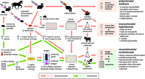
Figure 2. Biocompare.com search for different research antibody products, as of February 2024. The search engine could not distinguish between different products derived from the same original antibody preparation, e.g., with different labels or packaging sizes. As different labels are more commonly offered for secondary antibodies, which are mostly polyclonals, this segment may be slightly overrepresented.
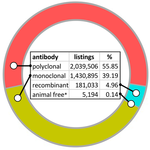
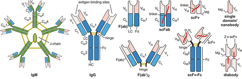
Figure 3. Antibody formats most frequently used in research assays: IgM, Immunoglobulin M; IgG, Immunoglobulin G; F(ab’)2, divalent fragment antigen binding; F(ab), fragment antigen binding, now usually named Fab; scFab, single chain fragment antigen binding; scFv-Fc, single chain fragment variable::Fc fusion protein; scFv, single chain fragment variable; diabody, dimerized scFv; and single domain antibodies/nanobody. Constant regions are colored dark blue (γ chains), green (µ chains), or light blue (κ or λ chains); variable (antigen binding) regions in pink. Yellow lines indicate disulfide bonds, red lines peptide linker, purple lines peptide tags. C, constant region, V, variable region, HC, heavy chain; LC light chain. Elements are not drawn to scale.